
Page 178 - Veterinary Histology of Domestic Mammals and Birds, 5th Edition
P. 178
160 Veterinary Histology of Domestic Mammals and Birds
Lymphocytes leave the lymph node by the lympho- pedunculated bursa is integrated into the dorsal wall of
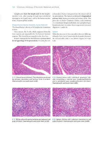
VetBooks.ir vascular route, after passing through inter-endothelial the proctodeum. The bursa is subdivided by longitudinal
openings in the lymph sinus, and by the haemovascular primary folds bearing secondary and tertiary folds. This
gives rise to distinct lymphatic lobules, each consisting
route, via postcapillary venules.
of a central pars lymphoepithelialis and a peripheral pars
Cloacal bursa (bursa cloacalis, bursa Fabricii) lymphoreticularis (in ratites this arrangement is reversed)
The cloacal bursa is the site of B-lymphocyte maturation (Figures 8.17 and 8.18).
in birds.
Once mature, the B cells, which originated from the Spleen
bone marrow, are responsible for the humoral immune While the structure of the avian spleen does not differ sig-
response. The cloacal bursa is specific to the class Aves. nificantly from that of mammals, the boundary between
In most avian species the cloacal bursa is a pedunculated the red and white pulp is less distinct (Figures 8.19 and
dorsal appendage of the proctodeum. In ratites, the non- 8.20).
8.17 Cloacal bursa (chicken). The lobulation produced 8.18 Cloacal bursa with individual lymphatic lob-
by primary, secondary and tertiary folds is evident. ules comprising a central pars lymphoepithelialis
Haematoxylin and eosin stain (x20). and a peripheral pars lymphoreticularis (chicken).
Haematoxylin and eosin stain (x80).
8.19 White pulp with central arteries and adjacent red 8.20 Splenic follicle with indistinct transition to red
pulp (chicken). Haematoxylin and eosin stain (x120). pulp (chicken). Haematoxylin and eosin stain (x240).
Vet Histology.indb 160 16/07/2019 14:59