
Page 177 - Veterinary Histology of Domestic Mammals and Birds, 5th Edition
P. 177
Immune system and lymphatic organs (organa Iymphopoetica) 159
erythrocytes, leucocytes, thrombocytes and macrophages · lymph heart (cor lymphaticum),
VetBooks.ir (Fig 8.15). Distributed throughout the reticular meshwork · mural lymphoreticular formations,
are irregularly expanded anastomosing vascular spaces
· a paucity of lymph nodes, limited to certain loca-
(diameter 12–50 μm) (splenic sinuses) (Figures 8.14 and
tions and
8.15). In some species these are not true sinuses, instead · the cloacal bursa (bursa Fabricii).
resembling venules (‘non-sinusal spleens’). The walls of
the sinuses are interrupted by longitudinally oriented
spaces that permit the passage of blood cells. Actively Lymph hearts and mural lymphoreticular formations are
phagocytosing macrophages tend to accumulate at these described in Chapter 6, ‘Circulatory system’.
sites.
The distribution of blood vessels in the spleen is char- Lymph nodes
acteristic for this organ (Figure 8.15). Arteries entering The relatively simple structure of avian lymph nodes
at the hilus divide into trabecular arteries. These pass resembles that of mural lymphoreticular formations
into the parenchyma as arteries of the white pulp, or (widely distributed specialisations of the walls of lymph
central arteries. The central arteries are surrounded vessels) and thus differs considerably from that of mam-
for some distance by a sleeve of lymphatic tissue, the malian lymph nodes.
periarteriolar lymphatic sheath (PALS). Branches of the Lymph nodes are found only in water and marsh birds,
arteries also enter the follicles. Some of these branches occurring in the neck (cervicothoracic lymph nodes) and
terminate in the marginal zone while others pass into the pelvic region (pelvic lymph nodes).
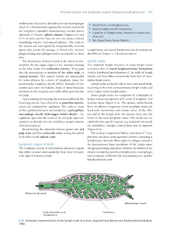
red pulp. Avian lymph nodes are composed of a labyrinth of
Upon entering the red pulp, the arteries exhibit tuft-like lymph sinuses interspersed with cords of lymphatic and
branching and are thus referred to as penicillar arteries, reticular tissue (Figure 8.16). The sinuses, which branch
which are continued by capillaries. The walls of some from the afferent component of the lymphatic vessel, are
of the capillaries become surrounded by a pericapillary lined with endothelium and contain valves. At the effer-
macrophage sheath (Schweigger–Seidel sheath). The ent end of the lymph node the sinuses drain into the
capillaries open into the cords of the red pulp (open cir- lumen of the same lymphatic vessel. The sinuses are not
culation) or directly into the medullary sinuses/venules subdivided into specific regions (e.g. marginal, intermedi-
(closed circulation). ate, medullary), though a central sinus may be apparent
Blood leaving the sinusoids/venules passes into red (Figure 8.16).
pulp veins and then trabecular veins, exiting the spleen The cords are composed of diffuse collections of T lym-
at the hilus via the splenic veins. phocytes and areas (avian germinal centres) containing B
lymphocytes. Reticular fibres (type III collagen) extend to
Lymphatic organs of birds the discontinuous basal membrane of the lymph sinus.
The lymphatic system of birds includes distinctive organs Antigen processing takes place between the lumina of the
that differ, in some cases markedly, from those of mam- sinuses (containing numerous lymphocytes, macrophages
mals. Special features include: and occasional red blood cells and granulocytes) and the
lymphoreticular cords.
8.16 Schematic representation of the lymph node of a duck, adapted from Berens von Rautenfeld and Budras,
1983.
Vet Histology.indb 159 16/07/2019 14:59