
Page 225 - Veterinary Histology of Domestic Mammals and Birds, 5th Edition
P. 225
Digestive system (apparatus digestorius) 207
olism, by the mucosa of the forestomach, particularly the The internal surface of the forestomach is lined by non-
VetBooks.ir rumen, and transferred to the bloodstream. In the oma- glandular mucosa (pars non-glandularis) (Figures
sum, the gruel-like ingesta are mechanically compressed 10.39 to 10.42 and Table 10.3). The papillated stratified
and water is absorbed. The forestomach constitutes the squamous epithelium is keratinised depending on the
non-glandular portion of the multi-chambered ruminant functional demands on the mucosa. In the tela submu-
stomach. Enzymatic digestion of the liquid portion of the cosa, branching blood vessels give off arterioles that lead
ingesta occurs in the glandular abomasum. The mucosa to subepithelial capillary networks, which drain into post-
of the abomasum represents the glandular portion of capillary venules. Veins merge in the tela submucosa and
the ruminant stomach. The forestomach consists of three course through the muscle layer towards the exterior.
compartments: As is typical for the musculomembranous gastrointes-
tinal tract, lymphatic vessels and autonomic intramural
· reticulum, ganglia are present in the tela submucosa. External to
· rumen and the tela submucosa is a tunica muscularis and a tunica
· omasum. serosa.
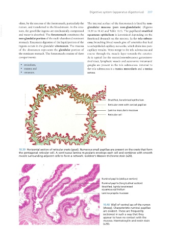
10.39 Horizontal section of reticular crests (goat). Numerous small papillae are present on the crests that form
the pentagonal reticular cell. A continuous lamina muscularis envelops each cell and combines with smooth
muscle surrounding adjacent cells to form a network. Goldner’s Masson trichrome stain (x20).
10.40 Wall of ventral sac of the rumen
(sheep). Characteristic ruminal papillae
are evident. These are frequently
sectioned in such a way that they
appear to have no contact with the
mucosa. Haematoxylin and eosin stain
(x20).
Vet Histology.indb 207 16/07/2019 15:01