Page 230 - Veterinary Histology of Domestic Mammals and Birds, 5th Edition
P. 230
212  Veterinary Histology of Domestic Mammals and Birds


       VetBooks.ir


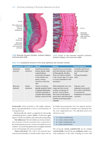
                   10.46  Muscular stomach (chicken). Goldner’s Masson   10.47  Cuticle of the muscular stomach (chicken).
                   trichrome stain (x20).                         Goldner’s Masson trichrome stain (x40).


                   Table 10.4  Comparative structure of the avian glandular and muscular stomach.

                    Compartment   Epithelium  Glands              Muscle                   Special features
                    Glandular     Simple      Superficial and deep   Lamina muscularis mucosae   Cuboidal epithelium,
                    stomach       columnar    tubular glands, deep   protrudes between clusters   secretes pepsinogen
                                              proprial glands     of deep glands, dividing   and
                                              extensively lobulated   these into distinct lobules;   HCl (oxynticopeptic
                                              in the chicken and   inner circular layer of muscle  cells)
                                              goose, mucous       in tunica muscularis is well
                                              secretion; endocrine   developed
                                              cells
                    Muscular      Simple      Clusters of tubular   Wall subdivided into two   Together with
                    stomach       columnar    glands; secretion forms  major and two minor   ingested stones (grit),
                                              a rigid grinding plate   muscles composed of   the cuticle contributes
                                              (cuticle) composed of   smooth muscle fibres;   to mechanical
                                              a keratin-like complex   the inner muscle layer is   breakdown of stomach
                                              (koilin); endocrine cells  particularly well developed;  contents
                                                                  tendinous centres present
                                                                  laterally


                   horizontalis (softer secretions of the simple columnar  be divided macroscopically into four separate muscles.
                   gastric mucosal epithelium) to form a continuous layer of  The inner circular layer is usually more developed than
                   varying thickness.                             the outer longitudinal layer. The individual muscles are
                      Biochemically, the cuticle is composed of a keratin-like  referred to as the:
                   carbohydrate-protein complex (koilin). Small stones (grit)
                   ingested with the food further assist with mechanical grinding.  ·  m. crassus caudodorsalis,
                      Tela submucosa: The tela submucosa forms an immo-  ·  m. crassus cranioventralis,
                   bile connection with the connective tissue components of   ·  m. tenuis craniodorsalis and
                   the lamina propria mucosae (a lamina muscularis mucosae   ·  m. tenuis caudoventralis.
                   is absent). As a result, the gastric mucosa is tightly anchored
                   to the overlying layer, the tunica muscularis.  The strong  m. crassus caudodorsalis and  m. crassus
                      Tunica muscularis: The wall of the muscular layer  cranioventralis extend from one tendinous centre (cen-
                   consists predominantly of smooth muscle tissue that can  trum tendineum) to the other, forming the dorsal and


       Vet Histology.indb   212                                                                                  16/07/2019   15:01

