Page 231 - Veterinary Histology of Domestic Mammals and Birds, 5th Edition
P. 231
Digestive system (apparatus digestorius)   213


                  ventral borders of the ventriculus. Lying between the thick  roscopic appearance of these components (for more detail
       VetBooks.ir  muscles are the weaker m. tenuis craniodorsalis and m.  see anatomy textbooks).
                  tenuis caudoventralis. In grain feeders, the ventriculus is
                                                                    The function of the small intestinal mucosa, supported
                                                                 by the activity of the muscular layers, is digestion of nutri-
                  the site of mechanical breakdown of food. Further diges-
                  tion of protein also occurs in this portion of the stomach.  ents and absorption of the resulting breakdown products.
                  The cuticle terminates at the ostium pyloricum. The   For the most part, the digestive processes that com-
                  mucosa of the pylorus contains pyloric glands (glandulae  mence in the proximal portion of the gastrointestinal tract
                  pyloricae) and numerous endocrine cells responsible for  are completed by reactions occurring in the small intestine.
                  local hormone production (e.g. gastrin cells, somatostatin  Further digestion takes place in the large intestine (within
                  cells).                                        fermentation chambers in the pig and horse). Ruminants
                     The histological features of the wall of the avian glan-  represent a special case, due to the role played in digestion
                  dular and muscular stomachs are listed in Table 10.4.  by the forestomach.
                                                                    Breakdown of nutrients is completed by the combined
                  Small intestine (intestinum tenue)             action of gastric juice (pepsin) and secretions of the intestinal
                  The small intestine comprises three structurally and func-  glands (including 1,6-glucosidase, aminopeptidase, dipepti-
                  tionally distinct segments, the duodenum, the jejunum and  dases and phosphodiesterases) and the pancreas (including
                  the ileum. Considerable species variation exists in the mac-  lipase, α-amylase, trypsin, chymotrypsin, carboxypeptidase,


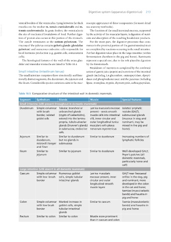
                  Table 10.5  Comparative structure of the intestinal wall in domestic mammals.
                   Segment     Epithelium     Glands                Muscle                Special features
                   Small intestine (villi present throughout in domestic mammals)
                   Duodenum    Simple columnar  Tubular, branched or   Lamina muscularis mucosae  Isolated lymphatic
                               with brush     unbranched glands     present – sends smooth   nodules (GALT),
                               border, isolated   (crypts of Lieberkühn),   muscle cells into intestinal  submucosal glands
                               goblet cells   extend into the lamina   villi, inner circular and   (mucous in dog and
                                              propria, tubulo-alveolar   outer longitudinal tunica   ruminant, may be
                                              glands (Brunner’s glands)  muscularis with plexus   mixed in the pig and
                                              in submucosa, endocrine  nervorum myentericus  horse).
                                              cells
                   Jejunum     Similar to     Similar to duodenum   Similar to duodenum   Increasing numbers of
                               duodenum,      but no glands in                            lymphatic follicles
                               microvilli longer  submucosa
                               and finer
                   Ileum       Similar to     Similar to jejunum    Similar to duodenum   Well-developed GALT,
                               jejunum                                                    Peyer’s patches (all
                                                                                          domestic mammals,
                                                                                          particularly horse and
                                                                                          calf)
                   Large intestine (villi absent in all domestic mammals)
                   Caecum      Simple columnar  Numerous goblet     Lamina muscularis     GALT near ileocaecal
                               with low  brush   cells, simple tubular   mucosae present, inner   orifice in the dog, pig
                               border         intestinal glands     circular and outer    and ruminant, more
                                                                    longitudinal smooth   developed in the colon
                                                                    muscle layers         in the cat and horse;
                                                                                          taeniae (musculoelastic
                                                                                          bands) and haustra in
                                                                                          pig and horse
                   Colon       Simple columnar  Marked increase in   Similar to caecum    Taenia (musculoelastic
                               with low brush   goblet cells, simple                      bands) and haustra in
                               border         tubular intestinal                          pig and horse
                                              glands
                   Rectum      Similar to colon  Similar to colon   Muscle more prominent
                                                                    than in caecum and colon


       Vet Histology.indb   213                                                                                  16/07/2019   15:01

