
Page 229 - Veterinary Histology of Domestic Mammals and Birds, 5th Edition
P. 229
Digestive system (apparatus digestorius) 211
GLANDULAR STOMACH (PROVENTRICULUS, PARS The loose connective tissue of the tela submucosa
VetBooks.ir The proventriculus continues from the oesophagus with- contains the submucosal nerve plexus (plexus nervorum
GLANDULARIS)
submucosus; Meissner plexus) and numerous vessels. In
out a clear anatomical boundary. In most species, the the tunica muscularis, a well-developed inner circular
mucosa is arranged in folds (Figures 10.44 and 10.45 and layer (stratum circulare) is surrounded by a thinner outer
Table 10.4). The mucosal surface is lined by mucus-secret- longitudinal layer (stratum longitudinale). The myenteric
ing simple columnar epithelium with microvilli. Tubular nerve plexus (plexus nervorum myentericus; Auerbach
glands situated in the lamina propria deliver their mucous plexus) lies between the two muscular layers. Externally,
secretory product onto the mucosal surface (Figures 10.44 the proventriculus is lined by a single-layered tunica serosa
and 10.45). These comprise: that is continuous with the intestinal peritoneal sac.
The proventriculus is typically separated from the
· superficial proventricular glands (glandulae proven- ventriculus by a narrow gastric isthmus (isthmus gastris).
triculares superficiales) and This transitional zone (zona intermedia) is usually free of
· deep proventricular glands (glandulae proventricu- mucosal elevations and folds and is devoid of glands. In
lares profundae). the chicken, the narrowing at the isthmus is attributable to
a high concentration of elastic fibres and a relatively thin
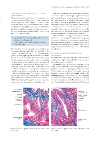
The superficial proventricular glands are simple tubu- muscular layer.
lar mucus-producing glands that open into folds in the
mucosa. The prominent layer of deep proventricular MUSCULAR STOMACH (VENTRICULUS, PARS
glands, or ‘deep proprial glands’, is bounded by a lamina MUSCULARIS)
muscularis mucosae. Portions of this muscle layer cleave Tunica mucosa: The gastric mucosa is irregularly subdi-
off and extend towards the surface, largely surrounding vided by folds (rugae ventriculi). These are absent near
the deep glands and protruding between the deep and the tendinous centres (see below).
more superficially located glands. While proventricular The luminal surface of the ventriculus is covered by a
glands are usually not lobed, distinct lobulation occurs tough, greenish-yellow layer known as the cuticle (cuticula
in chickens and geese. The openings of these glands are gastris) that acts akin to a grinding plate for the breakdown
located on mucosal elevations (papillae proventriculares). of food (Figures 10.46 and 10.47). The cuticle consists of
The cuboidal epithelium of the proventricular glands solidified secretions of clusters of tubular glands in the
secretes pepsinogen and HCl (oxynticopeptic cells). These lamina propria (glandulae ventriculares). Secretory prod-
cells contain numerous mitochondria and rough ER. uct is released onto the mucosal surface as compacted
Additional cell types produce gastric hormones (e.g. soma- cylinders, or columnae verticales, that harden to form
tostatin, bombesin and vasoactive intestinal peptide). rods. This solidified secretion combines with the matrix
10.44 Mucosa of glandular stomach (chicken). Azan 10.45 Mucosa of glandular stomach (chicken). Azan
stain (x20). stain (x40).
Vet Histology.indb 211 16/07/2019 15:01