
Page 292 - Veterinary Histology of Domestic Mammals and Birds, 5th Edition
P. 292
274 Veterinary Histology of Domestic Mammals and Birds
VetBooks.ir
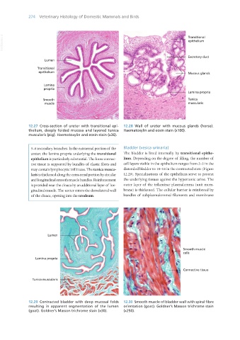
12.27 Cross-section of ureter with transitional epi- 12.28 Wall of ureter with mucous glands (horse).
thelium, deeply folded mucosa and layered tunica Haematoxylin and eosin stain (x100).
muscularis (pig). Haematoxylin and eosin stain (x36).
5–6 secondary branches. In the extrarenal portion of the Bladder (vesica urinaria)
ureter, the lamina propria underlying the transitional The bladder is lined internally by transitional epithe-
epithelium is particularly substantial. The loose connec- lium. Depending on the degree of filling, the number of
tive tissue is supported by bundles of elastic fibres and cell layers visible in the epithelium ranges from 2–3 in the
may contain lymphocytic infiltrates. The tunica muscu- distended bladder to 10–14 in the contracted state (Figure
laris is thickened along the extra-renal portion by circular 12.29). Specialisations of the epithelium serve to protect
and longitudinal smooth muscle bundles. Reinforcement the underlying tissues against the hypertonic urine. The
is provided near the cloaca by an additional layer of lon- outer layer of the trilaminar plasmalemma (unit mem-
gitudinal muscle. The ureter enters the dorsolateral wall brane) is thickened. The cellular barrier is reinforced by
of the cloaca, opening into the urodeum. bundles of subplasmalemmal filaments and membrane
12.29 Contracted bladder with deep mucosal folds 12.30 Smooth muscle of bladder wall with spiral fibre
resulting in apparent segmentation of the lumen orientation (goat). Goldner’s Masson trichrome stain
(goat). Goldner’s Masson trichrome stain (x30). (x250).
Vet Histology.indb 274 16/07/2019 15:04