
Page 309 - Veterinary Histology of Domestic Mammals and Birds, 5th Edition
P. 309
Male reproductive system (organa genitalia masculina) 291
VetBooks.ir ciliated
Stereo-
pseudo-
stratified
columnar
epithelium
Smooth
muscle
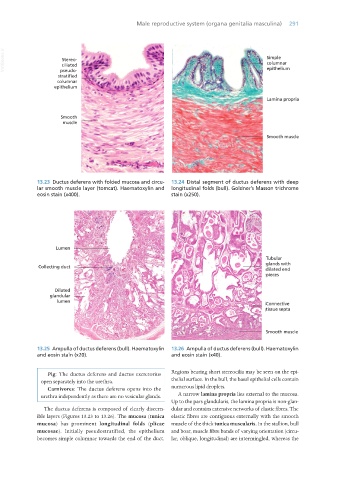
13.23 Ductus deferens with folded mucosa and circu- 13.24 Distal segment of ductus deferens with deep
lar smooth muscle layer (tomcat). Haematoxylin and longitudinal folds (bull). Goldner’s Masson trichrome
eosin stain (x400). stain (x250).
13.25 Ampulla of ductus deferens (bull). Haematoxylin 13.26 Ampulla of ductus deferens (bull). Haematoxylin
and eosin stain (x20). and eosin stain (x40).
Pig: The ductus deferens and ductus excretorius Regions bearing short stereocilia may be seen on the epi-
open separately into the urethra. thelial surface. In the bull, the basal epithelial cells contain
Carnivores: The ductus deferens opens into the numerous lipid droplets.
urethra independently as there are no vesicular glands. A narrow lamina propria lies external to the mucosa.
Up to the pars glandularis, the lamina propria is non-glan-
The ductus deferens is composed of clearly discern- dular and contains extensive networks of elastic fibres. The
ible layers (Figures 13.23 to 13.26). The mucosa (tunica elastic fibres are contiguous externally with the smooth
mucosa) has prominent longitudinal folds (plicae muscle of the thick tunica muscularis. In the stallion, bull
mucosae). Initially pseudostratified, the epithelium and boar, muscle fibre bands of varying orientation (circu-
becomes simple columnar towards the end of the duct. lar, oblique, longitudinal) are intermingled, whereas the
Vet Histology.indb 291 16/07/2019 15:05