
Page 368 - Veterinary Histology of Domestic Mammals and Birds, 5th Edition
P. 368
350 Veterinary Histology of Domestic Mammals and Birds
VetBooks.ir
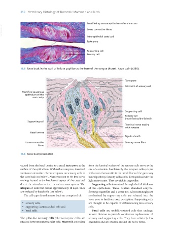
16.5 Taste buds in the wall of foliate papillae at the base of the tongue (horse). Azan stain (x250).
16.6 Taste bud (schematic).
extend from the basal lamina to a small taste pore at the from the luminal surface of the sensory cells serve as the
surface of the epithelium. Within the taste pore, dissolved site of excitation. Basolaterally, the receptor cells synapse
substances stimulate chemoreceptors on sensory cells in with axons that constitute the initial fibres of the gustatory
the taste bud (see below). Numerous (up to 50) free nerve neural pathway. Sensory cells can be distinguished with the
endings located at the basolateral aspect of the taste bud light microscope. They are rich in organelles.
direct the stimulus to the central nervous system. The Supporting cells also extend through the full thickness
lifespan of taste bud cells is approximately 10 days. They of the epithelium. These contain abundant enzyme-
are replaced by basal cells (see below). forming organelles and a dense ER. Glycosaminoglycans
The cell types found in taste buds are comprised of: synthesised by supporting cells are released into the
taste pore to facilitate taste perception. Supporting cells
· sensory cells, are thought to be capable of differentiating into sensory
· supporting (sustentacular) cells and cells.
· basal cells. Basal cells are undifferentiated cells that undergo
mitotic division to provide continuous replacement of
The pillar-like sensory cells (chemoreceptor cells) are sensory and supporting cells. They have relatively few
situated between sustentacular cells. Microvilli extending organelles and are situated around the nerve fibres.
Vet Histology.indb 350 16/07/2019 15:07