
Page 371 - Veterinary Histology of Domestic Mammals and Birds, 5th Edition
P. 371
Receptors and sense organs (organa sensuum) 353
epithelium, supporting cells perform a role equivalent to within the eye and the associated neural tracts are formed
VetBooks.ir that of glial cells. In addition, these organelle-rich cells from the diencephalon during embryonic development.
exocytose mucoid substances. Intracellular pigments give
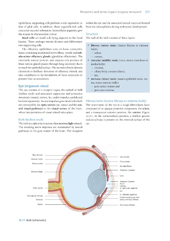
Structure
the mucus its characteristic colour.
Basal cells are small cells lying adjacent to the basal The wall of the bulb consists of three layers:
lamina. These undergo mitotic division and differentiate
into supporting cells. · fibrous (outer) tunic (tunica fibrosa or externa
The olfactory epithelium rests on loose connective bulbi):
tissue containing myelinated nerve fibres, vessels and tub- − sclera,
ulo-acinar olfactory glands (glandulae olfactoriae). The − cornea,
extremely watery protein- and enzyme-rich product of · vascular (middle) tunic (uvea, tunica vasculosa or
these serous glands passes through long excretory ducts media bulbi):
to reach the epithelial surface. The secretion binds odorant − choroid,
chemicals to facilitate detection of olfactory stimuli, and − ciliary body (corpus ciliare),
also contributes to the breakdown of these molecules to − iris,
prevent their accumulation. · nervous (inner) tunic (neuro-epithelial tunic, ret-
ina, tunica interna bulbi):
Eye (organum visus) − pars optica retinae and
The eye consists of a receptor organ, the eyeball or bulb − pars caeca retinae.
(bulbus oculi) and associated supportive and protective
structures (vessels, nerves, fat, ocular muscles, eyelids and
lacrimal apparatus). Neural impulses generated in the bulb Fibrous tunic (tunica fibrosa or externa bulbi)
are conveyed by the optic nerves (nn. optici) and the cen- The outer tunic of the eye is a tough fibro-elastic layer
tral visual pathways to the visual cortex of the brain, composed of an opaque posterior component, the sclera,
where interpretation of visual stimuli takes place. and a transparent anterior portion, the cornea (Figure
16.11). At the corneoscleral junction, a shallow groove
Bulb (bulbus oculi) (sulcus sclerae) is present on the external surface of the
The bulb is a spheroid structure that receives light stimuli. eye.
The resulting nerve impulses are transmitted by neural
pathways to the grey matter of the brain. The receptors
Neural tunic
Ora serrata
Vascular tunic
Ciliary body
Fibrous tunic
Zonular fibres
Posterior chamber
Vitreous body
Lens
Anterior chamber
Pupil
Cornea
Optic nerve m. sphincter pupillae
Iris
m. dilatator pupillae
Pars optica retinae
Iridocorneal angle with
Choroid plexus venosus sclerae
Sclera Pars caeca retinae
16.11 Bulb (schematic).
Vet Histology.indb 353 16/07/2019 15:07