
Page 376 - Veterinary Histology of Domestic Mammals and Birds, 5th Edition
P. 376
358 Veterinary Histology of Domestic Mammals and Birds
species, also the iris sphincter muscle. Parasympathetic
The striated pupillary muscle of birds is under
VetBooks.ir voluntary control and, in contrast to mammals, is impulses originating primarily from the oculomotor nerve
are conveyed by postganglionic fibres to the m. sphincter
not responsive to commonly used ophthalmic drugs.
pupillae and the ciliary muscle. The effects of parasym-
Thus, while parasympatholytics are routinely used in
mammalian patients to achieve mydriasis for ophthal- pathetic stimulation include pupillary constriction and
moscopic examination of the fundus, this practice is contraction of the ciliary muscle for accommodation.
ineffective in birds. In several diving birds, including Parasympathetic signals are transmitted by acetylcho-
penguins and cormorants, the m. sphincter pupillae line secreted at the neuro-effector junction. Cholinergic
is particularly well developed and the lens protrudes agents (e.g. pilocarpine, carbachol) activate muscarinic
through the pupil during accommodation. This receptors, resulting in constriction of the pupil and contrac-
increases the refractive capacity of the lens, compen- tion of the ciliary muscle. Blockage of these nerve impulses
sating for reduced corneal refraction under water. by the plant alkaloids atropine and hyoscine causes passive
mydriasis and disruption of accommodation.
The sympathetic (adrenergic) system is activated by
INNERVATION OF THE IRIS AND CILIARY BODY noradrenaline, serving as a neurotransmitter, and by cir-
The muscular and vascular components of the mammalian culating adrenaline. The effect of these molecules, which
iris are controlled by neural and endocrine mechanisms. bind with α- and β-receptors, can be modified by catecho-
The iris and ciliary body receive sympathetic and para- lamine-depleting agents (e.g. reserpine).
sympathetic innervation from the short and long ciliary Other substances that act as neurotransmitters in the
nerves. Postganglionic sympathetic fibres supply the m. regulation of iris and ciliary muscle function include the
dilatator pupillae, the blood vessels of the iris and ciliary neuropeptides substance P, vasoactive intestinal peptide
processes, stromal melanocytes, fibroblasts and, in some (VIP) and neuropeptide Y (NPY).
Neural tunic (retina, tunica interna bulbi)
The retina is divided into an anterior portion devoid of
photoreceptors, the pars caeca retinae, and a posterior
photosensitive portion, the pars optica retinae (Figure
16.11). Both portions consist of an inner and outer layer,
reflecting their embryonic origin.
In the pars caeca retinae, the two leaves of the optic
cup remain largely undifferentiated, each forming a simple
layer of epithelium. The two layers are closely apposed
but remain separate. Based on its relationship to the ciliary
body and iris, the pars caeca retinae is subdivided into the:
· pars ciliaris retinae,
· pars iridica retinae and
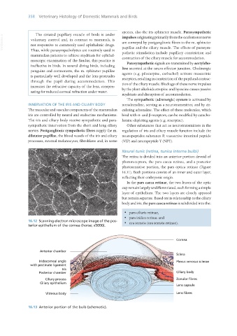
16.12 Scanning electron microscope image of the pos- · ora serrata (ora serrata retinae).
terior epithelium of the cornea (horse; x5000).
16.13 Anterior portion of the bulb (schematic).
Vet Histology.indb 358 16/07/2019 15:07