
Page 378 - Veterinary Histology of Domestic Mammals and Birds, 5th Edition
P. 378
360 Veterinary Histology of Domestic Mammals and Birds
VetBooks.ir Discontinuous
cellular layer
Stroma with melanocytes
m. dilatator pupillae
Pigmented portion
of pars iridica retinae
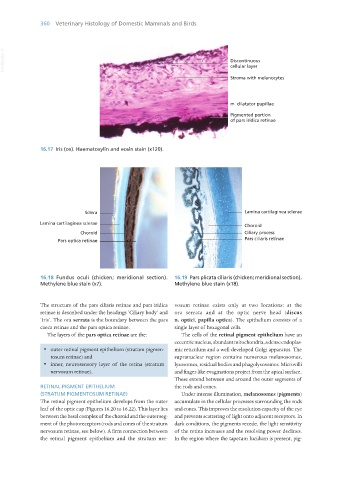
16.17 Iris (ox). Haematoxylin and eosin stain (x120).
16.18 Fundus oculi (chicken; meridional section). 16.19 Pars plicata ciliaris (chicken; meridional section).
Methylene blue stain (x7). Methylene blue stain (x18).
The structure of the pars ciliaris retinae and pars iridica vosum retinae exists only at two locations: at the
retinae is described under the headings ‘Ciliary body’ and ora serrata and at the optic nerve head (discus
‘Iris’. The ora serrata is the boundary between the pars n. optici, papilla optica). The epithelium consists of a
caeca retinae and the pars optica retinae. single layer of hexagonal cells.
The layers of the pars optica retinae are the: The cells of the retinal pigment epithelium have an
eccentric nucleus, abundant mitochondria, a dense endoplas-
· outer retinal pigment epithelium (stratum pigmen- mic reticulum and a well-developed Golgi apparatus. The
tosum retinae) and supranuclear region contains numerous melanosomes,
· inner, neurosensory layer of the retina (stratum lysosomes, residual bodies and phagolysosomes. Microvilli
nervosum retinae). and finger-like evaginations project from the apical surface.
These extend between and around the outer segments of
RETINAL PIGMENT EPITHELIUM the rods and cones.
(STRATUM PIGMENTOSUM RETINAE) Under intense illumination, melanosomes (pigments)
The retinal pigment epithelium develops from the outer accumulate in the cellular processes surrounding the rods
leaf of the optic cup (Figures 16.20 to 16.22). This layer lies and cones. This improves the resolution capacity of the eye
between the basal complex of the choroid and the outer seg- and prevents scattering of light onto adjacent receptors. In
ment of the photoreceptors (rods and cones of the stratum dark conditions, the pigments recede, the light sensitivity
nervosum retinae, see below). A firm connection between of the retina increases and the resolving power declines.
the retinal pigment epithelium and the stratum ner- In the region where the tapetum lucidum is present, pig-
Vet Histology.indb 360 16/07/2019 15:07