
Page 382 - Veterinary Histology of Domestic Mammals and Birds, 5th Edition
P. 382
364 Veterinary Histology of Domestic Mammals and Birds
membrane constitutes the boundary between the retina
VetBooks.ir and the vitreous body.
Müller cells extend perpendicularly through the ret-
ina. Their nuclei form part of the inner nuclear layer. In
the outer portion of the cell, the cytoplasm narrows and
extends between the photoreceptors to the outer extrem-
ity of the inner segment. Zonulae adherentes between the
Müller cells give rise to the external limiting membrane
(stratum limitans gliae externum). Lying at the outer
aspect of the outer nucler layer (Figures 16.26 and 16.29),
the external limiting membrane forms an important meta-
bolic barrier between the rods and cones and the inner
layers of the retina. The more internal layers are supplied
by the retinal vessels, while nourishment of the photore-
ceptors occurs by diffusion of molecules across the retinal
pigment epithelium.
Area centralis rotunda: In primates, the area of great-
est visual acuity is a well-circumscribed yellowish region
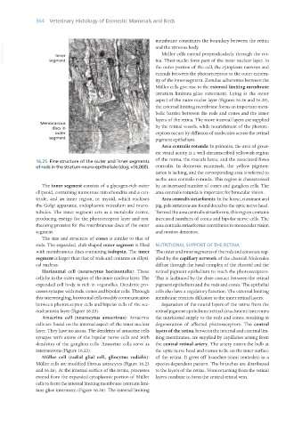
16.25 Fine structure of the outer and inner segments of the retina, the macula lutea, and the associated fovea
of rods in the stratum neuro-epitheliale (dog; x16,000). centralis. In domestic mammals, the yellow pigmen-
tation is lacking, and the corresponding area is referred to
as the area centralis rotunda. This region is characterised
The inner segment consists of a glycogen-rich outer by an increased number of cones and ganglion cells. The
ellipsoid, containing numerous mitochondria and a cen- area centralis rotunda is important for binocular vision.
triole, and an inner region, or myoid, which encloses Area centralis striaeformis: In the horse, ruminant and
the Golgi apparatus, endoplasmic reticulum and micro- pig, pale striations are found dorsal to the optic nerve head.
tubules. The inner segment acts as a metabolic centre, Termed the area centralis striaeformis, this region contains
producing energy for the photoreceptor layer and syn- increased numbers of cones and bipolar nerve cells. The
thesising proteins for the membranous discs of the outer area centralis striaeformis contributes to monocular vision
segment. and motion detection.
The size and structure of cones is similar to that of
rods. The expanded, club-shaped outer segment is filled NUTRITIONAL SUPPORT OF THE RETINA
with membranous discs containing iodopsin. The inner The outer and inner segments of the rods and cones are sup-
segment is larger than that of rods and contains an ellipti- plied by the capillary network of the choroid. Molecules
cal nucleus. diffuse through the basal complex of the choroid and the
Horizontal cell (neurocytus horizontalis): These retinal pigment epithelium to reach the photoreceptors.
cells lie in the outer region of the inner nuclear layer. The This is facilitated by the close contact between the retinal
expanded cell body is rich in organelles. Dendritic pro- pigment epithelium and the rods and cones. The epithelial
cesses synapse with rods, cones and bipolar cells. Through cells also have a regulatory function. The external limiting
this intermingling, horizontal cells modify communication membrane restricts diffusion to the inner retinal layers.
between photoreceptor cells and bipolar cells of the sec- Separation of the neural layers of the retina from the
ond neuron layer (Figure 16.23). retinal pigment epithelium (retinal detachment) interrupts
Amacrine cell (neurocytus amacrinus): Amacrine the nutritional supply to the rods and cones, resulting in
cells are found on the internal aspect of the inner nuclear degeneration of affected photoreceptors. The central
layer. They have no axons. The dendrites of amacrine cells layers of the retina, between the internal and external lim-
synapse with axons of the bipolar nerve cells and with iting membranes, are supplied by capillaries arising from
dendrites of the ganglion cells. Amacrine cells serve as the central retinal artery. The artery enters the bulb at
interneurons (Figure 16.23). the optic nerve head and comes to lie on the inner surface
Müller cell (radial glial cell, gliocytus radialis): of the retina. It gives off branches (rami centrales) in a
Müller cells are modified fibrous astrocytes (Figure 16.23 species-dependent pattern. The branches are distributed
and 16.26). At the internal surface of the retina, processes to the layers of the retina. Veins returning from the retinal
extend from the expanded cytoplasmic portion of Müller layers combine to form the central retinal vein.
cells to form the internal limiting membrane (stratum limi-
tans gliae internum) (Figure 16.26). The internal limiting
Vet Histology.indb 364 16/07/2019 15:07