
Page 54 - Veterinary Histology of Domestic Mammals and Birds, 5th Edition
P. 54
36 Veterinary Histology of Domestic Mammals and Birds
filaments attach at these locations, enhancing the strength
VetBooks.ir of the cell-to-cell contact (Figures 1.47 and 1.49). The
mechanical function of desmosomes is supported by the
ring-like zonula adherens.
Desmosomes are the sole form of anchoring junction
found in the stratified epithelia of the skin. In cuboidal
and columnar epithelia, desmosomes and zonula adher-
ens usually occur together. Desmosomes (diameter 0.4 ×
0.25 μm) do not surround the cell as a continuous struc-
ture, rather they form spot- or plaque-like connections
between neighbouring cells.
While they are found primarily in epithelia, they also
occur between cardiac muscle cells. The width of the
space between segments of plasma membrane joined by a
desmosome is 20–40 nm.
On the cytoplasmic side of desmosomes, various
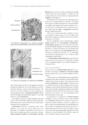
1.48 Electron micrograph of a zonula occludens plaque proteins (e.g. desmoplakin, plakoglobins,
adjoined by interdigitating cell processes (x21,000). desmocalmin) aggregate to form a dense structure (des-
mosomal attachment plaque) in which the intermediate
filaments are anchored. Between the two desmosomal
plaques is an integrated band (intermediate line) con-
2+
taining the extracellular portions of Ca -dependent
transmembrane glycoproteins of the cadherin family
(desmogleins, desmocollins).
The intermediate (keratin) filaments connected to the
desmosome also serve as part of the supportive cytoskel-
eton, extending throughout the cytoplasm along lines of
mechanical force.
GAP JUNCTION (NEXUS)
Gap junctions are communicating junctions that per-
mit direct exchange of chemical or electrical signals
between adjacent cells (e.g. in surface epithelia, smooth
muscle).
Gap junctions are widely distributed through tissues
and organs, particularly in epithelia, smooth muscle tissue,
1.49 Electron micrograph of a desmosome (x45,000). cardiac muscle and nervous tissue. Their diverse functions
include coordination of the activity of adjacent cells
sation and selective restriction of the passage of substances (e.g. epithelia involved in fluid transport and electrolyte
through the epithelium, the zonulae adherentes (and des- exchange, vascular and intestinal smooth muscle) and facil-
mosomes) perform a purely mechanical function. The itation of the passage of regulatory molecules and smaller
presence of zonula occludens and zonula adherens in metabolites between cells. The intercellular space in gap
close proximity gives rise to junctional complexes. When junctions is very narrow (2–5 nm), appearing electron
viewed with light microscopy, these appear as a network microscopically as a mere ‘gap’.
referred to as the terminal bar. Gap junctions are composed of channel proteins
Intercellular junctions in which membrane proteins arranged in a circle to form the wall of a pore. Referred
interact with intermediate filaments (keratin filaments) to as connexons, these protein complexes consist of six
include desmosomes (lateral cell membrane) and hemides- integral membrane subunits (connexins) configured to
mosomes (basal cell membrane). form a tunnel in the plasmalemma of each adjoining cell.
Desmosomes (Gk desmo = connection, soma = body) The connexons in apposing membranes line up to form a
(diameter 0.3–0.5 μm) are disc-like plates that serve channel that connects the two cells. The number of pores
primarily to strengthen intercellular connections. At (up to 2 nm wide, 20 nm long) within a gap junction varies
these sites, the apposing surface membranes condense. widely, up to several hundred. Their ability to open and
Intracytoplasmic bundles of intermediate (keratin) close reversibly is integral to their function.
Vet Histology.indb 36 16/07/2019 14:54