
Page 56 - Veterinary Histology of Domestic Mammals and Birds, 5th Edition
P. 56
38 Veterinary Histology of Domestic Mammals and Birds
Proteoglycans (heparin sulfate and chondroitin sul-
VetBooks.ir fate) form the bulk of the matrix, which, due to its anionic
nature, is heavily hydrated. The high proteoglycan content
plays an important role in regulation of the passage of ions
through the basal lamina. Both proteoglycans and laminin
are considered products of the epithelial cells.
Laminin contributes substantially to the integration
of the basal lamina with the base of the epithelium.
Entactin (nidogen) and fibronectin stabilise the basal
lamina and form part of the connection with the lamina
fibroreticularis.
The basal lamina has several functions. As well as
attaching the epithelium to the underlying connective tis-
sue, it serves to compartmentalise superficial tissue layers,
acting as a structural barrier between the epithelium (also
muscle and nerve tissue, see below) and the connective
tissue. In addition, the basal lamina functions as a selective
filter, regulating the transport of substances via integral
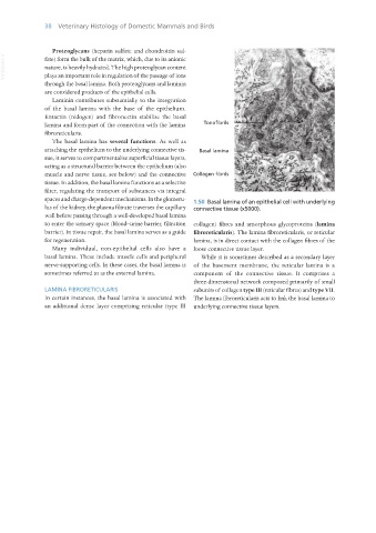
spaces and charge-dependent mechanisms. In the glomeru- 1.50 Basal lamina of an epithelial cell with underlying
lus of the kidney, the plasma filtrate traverses the capillary connective tissue (x5000).
wall before passing through a well-developed basal lamina
to enter the urinary space (blood–urine barrier, filtration collagen) fibres and amorphous glycoproteins (lamina
barrier). In tissue repair, the basal lamina serves as a guide fibroreticularis). The lamina fibroreticularis, or reticular
for regeneration. lamina, is in direct contact with the collagen fibres of the
Many individual, non-epithelial cells also have a loose connective tissue layer.
basal lamina. These include muscle cells and peripheral While it is sometimes described as a secondary layer
nerve-supporting cells. In these cases, the basal lamina is of the basement membrane, the reticular lamina is a
sometimes referred to as the external lamina. component of the connective tissue. It comprises a
three-dimensional network composed primarily of small
LAMINA FIBRORETICULARIS subunits of collagen type III (reticular fibres) and type VII.
In certain instances, the basal lamina is associated with The lamina fibroreticularis acts to link the basal lamina to
an additional dense layer comprising reticular (type III underlying connective tissue layers.
Vet Histology.indb 38 16/07/2019 14:54