
Page 76 - Veterinary Histology of Domestic Mammals and Birds, 5th Edition
P. 76
58 Veterinary Histology of Domestic Mammals and Birds
Serous glands (glandulae serosae) produce a watery secre- densely staining flattened nuclei lie at the base of the cell.
VetBooks.ir tion rich in protein and enzymes. The nuclei of the cells The cytoplasm has a foamy, vacuolated appearance, as the
of the secretory units are usually spherical and are located mucinogen (mucin precursor) within the cell is largely lost
centrally or slightly towards the base of the cell. Secretory during histological processing (Figures 2.33, 2.47 and 2.48).
granules of serous glands have a dense centre and a paler Mucin stains pale blue with haematoxylin and eosin, and
rim. The cytoplasm of the secretory cells is acidophilic and bright magenta with the PAS technique. Mucin also stains
the lumen of the secretory unit is narrow. Other morpho- strongly with alcian blue, due to the high sialic acid and
logical cellular features include pronounced infolding of polysaccharide content of the mucin molecule. Mucin
the basement membrane and finger-like interdigitations is found in many glandular secretions, particularly those
between neighbouring cells. Intercellular channels (secre- of the salivary glands. It serves as both a protectant and
tory capillaries) bounded by zonulae occludentes may a lubricant. The protein component of the mucinogen
be present apically, permitting a transient increase in the molecule is produced by the rER, while the carbohydrate
secretory surface of the cell. Microvilli are present on the portion is synthesised by the Golgi apparatus. Mucinogen
luminal surface (Figures 2.33, 2.45 and 2.46). is stored within the cell in pro-secretory granules. On
Mucous glands (glandulae mucosae) produce mucin, a secretion into the lumen of the secretory unit, mucinogen
glycoprotein with side chains containing oligosaccharides is hydrated to form mucin.
(incorporating glucose, galactose, mannose, N-acetyl- Seromucous (mixed) glands (glandulae seromucosae)
hexosamine and sialic acid). The secretory cells of mucous contain both serous cells and mucous cells. The former
glands are usually arranged in a single layer and are pyram- produce a secretion high in protein; the latter secrete pro-
idal in shape, with a blunted point at their apical surface. teoglycans and mucin. The serous secretory units appear
The cell borders are well demarcated, and the lumen of to form half-moon-shaped caps (serous demilunes, cres-
the secretory unit is relatively wide. Typically, basophilic, cents of Gianuzzi) around the mucous units. However,
Table 2.2 Structural differences between serous and mucous gland cells.
Characteristic Serous gland cell Mucous gland cell
Shape of cell Wedge-shaped to pyramidal, broader at base than Columnar or pyramidal
apex
Shape and Spherical (round in cross-section), lightly staining, Flattened, densely staining, at
position of central to basal half of cell margin of cell base
nucleus
Staining of Haematoxylin and eosin: basophilic (blue) in basal Basophilic in basal region, pale in
cytoplasm region, eosinophilic (red) apically supranuclear region
Function Protein synthesis Mucin synthesis
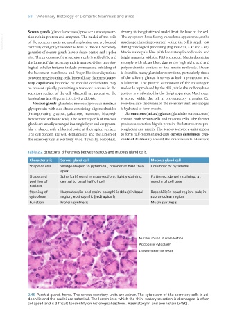
2.45 Parotid gland, horse. The serous secretory units are acinar. The cytoplasm of the secretory cells is aci-
dophilic and the nuclei are spherical. The lumen into which the thin, watery secretion is discharged is often
collapsed and is difficult to identify on histological sections. Haematoxylin and eosin stain (x480).
Vet Histology.indb 58 16/07/2019 14:55