
Page 77 - Veterinary Histology of Domestic Mammals and Birds, 5th Edition
P. 77
Epithelial tissue (textus epithelialis) 59
VetBooks.ir
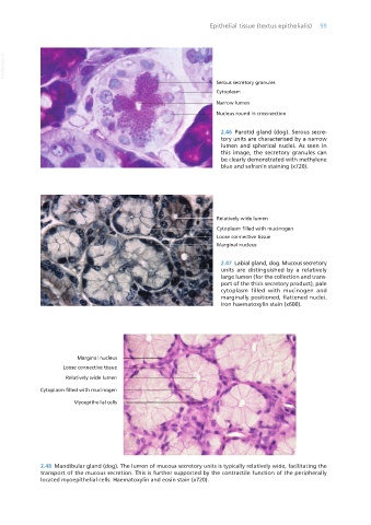
Serous secretory granules
Cytoplasm
Narrow lumen
Nucleus round in cross-section
2.46 Parotid gland (dog). Serous secre-
tory units are characterised by a narrow
lumen and spherical nuclei. As seen in
this image, the secretory granules can
be clearly demonstrated with methylene
blue and safranin staining (x720).
2.47 Labial gland, dog. Mucous secretory
units are distinguished by a relatively
large lumen (for the collection and trans-
port of the thick secretory product), pale
cytoplasm filled with mucinogen and
marginally positioned, flattened nuclei.
Iron haematoxylin stain (x600).
2.48 Mandibular gland (dog). The lumen of mucous secretory units is typically relatively wide, facilitating the
transport of the mucous secretion. This is further supported by the contractile function of the peripherally
located myoepithelial cells. Haematoxylin and eosin stain (x720).
Vet Histology.indb 59 16/07/2019 14:55