
Page 309 - The Veterinary Care of the Horse
P. 309
The procedure for scanning
The legs are clipped and scrubbed, then covered with a special gel. Modern ultrasound
VetBooks.ir machines produce very good quality, detailed images. Longitudinal and transverse images are
taken at several locations between the knee and the fetlock. The signs of tendon damage
include:
• an increase in cross-sectional area of the tendon
• area(s) of decreased density of the tendon structure – these show as dark areas within the
tendon (Figure 7.3b)
• change in shape of the tendon
• loss of longitudinal alignment of the fibres on the longitudinal view.
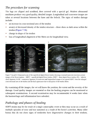
Figure 7.3a and b Ultrasound scans of the superficial digital flexor tendon showing a) normal transverse (sections across)
images of the flexor tendons – SDFT = superficial digital flexor tendon, DDFT = deep digital flexor tendon, ICL = inferior
check ligament, SUSP = suspensory ligament; b) transverse and longitudinal images of an injured SDFT. Note the enlarged
cross-sectional area of the tendon and the dark appearance due to disruption of the tendon fibres.
By examining all the images, the vet will know the position, the extent and the severity of the
damage. Good quality images are essential so that the healing progress can be monitored at
subsequent examinations. A second examination may be recommended 4 weeks later when
the haemorrhage and inflammation have subsided.
Pathology and phases of healing
SDFT strains may be the result of a single catastrophic event or they may occur as a result of
multiple episodes of wear and tear sustained as a result of the horse’s activities. Many older
horses that do not show signs of tendonitis have degenerative changes in their tendons.