
Page 13 - GP fall 2023
P. 13
At this time, a panoramic radiograph was taken, which revealed a The patient was given 300 mg of clindamycin immediately due
large periapical radiolucency centered on the distal root of #31 but to a poorly documented history of penicillin sensitivity, and the
encompassing both roots (Figure 4). The lesion measured 10 mm extraction was carried out without complications. During the
in diameter and overlapped the inferior mandibular canal. extraction, the socket was irrigated with saline until clear and
inspected for extension of the infection. The inferior man-
dibular nerve was observed, and 0.5 cc of osseous graft was
placed with a resorbable membrane for socket preservation.
The patient was released into the care of her physician with
the alert for further investigation of Ludwig’s angina. The
antibiotic course was completed, and the patient reported no
symptoms on follow-up appointments.
Discussion
While the presentation of mandibular teeth with periapical
radiolucency is a common occurrence in dental practice, it is
often imaged only with standard 2-dimensional views. Fur-
ther, patients are not often asked to reflect on neck, esophagus,
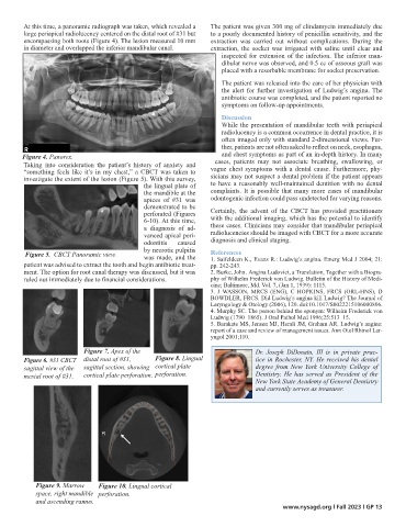
Figure 4. Panorex. and chest symptoms as part of an in-depth history. In many
Taking into consideration the patient’s history of anxiety and cases, patients may not associate breathing, swallowing, or
Figure 4. Panorex.
“something feels like it’s in my chest,” a CBCT was taken to vague chest symptoms with a dental cause. Furthermore, phy-
Figure 4. Panorex.
investigate the extent of the lesion (Figure 5). With this survey, sicians may not suspect a dental problem if the patient appears
the lingual plate of
Figure 4. Panorex. to have a reasonably well-maintained dentition with no dental
complaints. It is possible that many more cases of mandibular
the mandible at the
apices of #31 was odontogenic infection could pass undetected for varying reasons.
demonstrated to be
perforated (Figures Certainly, the advent of the CBCT has provided practitioners
6-10). At this time, with the additional imaging, which has the potential to identify
these cases. Clinicians may consider that mandibular periapical
a diagnosis of ad-
Figure 7. Apex of the distal root of #31, sagittal section, showing cortical plate perforation.
radiolucencies should be imaged with CBCT for a more accurate
vanced apical peri-
odontitis caused diagnosis and clinical staging.
by necrotic pulpitis
Figure 5. CBCT Panoramic view. References
Figure 5. CBCT Panoramic view. was made, and the 1. Saifeldeen K., Evans R.: Ludwig’s angina. Emerg Med J 2004; 21:
patient was advised to extract the tooth and begin antibiotic treat- pp. 242-243.
ment. The option for root canal therapy was discussed, but it was 2. Burke, John. Angina Ludovici, a Translation, Together with a Biogra-
ruled out immediately due to financial considerations.
phy of Wilhelm Frederick von Ludwig. Bulletin of the History of Medi-
Figure 5. CBCT Panoramic view. Figure 7. Apex of the distal root of #31, sagittal section, showing cortical plate perforation.
cine; Baltimore, Md. Vol. 7, (Jan 1, 1939): 1115.
3. J WASSON, MRCS (ENG), C HOPKINS, FRCS (ORL-HNS), D
BOWDLER, FRCS. Did Ludwig’s angina kill Ludwig? The Journal of
Laryngology & Otology (2006), 120. doi:10.1017/S0022215106000806.
4. Murphy SC. The person behind the eponym: Wilhelm Frederick von
Ludwig (1790–1865). J Oral Pathol Med 1996;25:513–15.
Figure 5. CBCT Panoramic view. 5. Barakate MS, Jensen MJ, Hemli JM, Graham AR. Ludwig’s angina:
report of a case and review of management issues. Ann Otol Rhinol Lar-
Figure 6. #31 CBCT sagittal view of the mesial root of #31. yngol 2001;110.
Figure 7. Apex of the Dr. Joseph DiDonato, III is in private prac-
Figure 6. #31 CBCT distal root of #31, Figure 8. Lingual tice in Rochester, NY. He received his dental
sagittal view of the sagittal section, showing cortical plate
Figure 7. Apex of the distal root of #31, sagittal section, showing cortical plate perforation.
Fig 8 Lingual cortical plate perforation degree from New York University College of
Figure 6. #31 CBCT sagittal view of the mesial root of #31.
Fig 8 Lingual cortical plate perforation
mesial root of #31.
cortical plate perforation. perforation. Dentistry. He has served as President of the
New York State Academy of General Dentistry
and currently serves as treasurer.
R
Fig 8 Lingual cortical plate perforation
Figure 6. #31 CBCT sagittal view of the mesial root of #31.
Fig 9 marrow space right mandible and ascending ramus
Figure 9. Marrow Figure 10. Lingual cortical Fig 10 Lingual cortical perforation
space, right mandible perforation.
and ascending ramus.
www.nysagd.org l Fall 2023 l GP 13
Fig 9 marrow space right mandible and ascending ramus
Fig 9 marrow space right mandible and ascending ramus