
Page 16 - GP Fall 2019
P. 16
Root of the Problem: A Case Report on
Unspecific Endodontically Involved Teeth
by Shariss Ostrager, Dr. Angela De Bartolo, Dr. Gene Sherwin, and Dr. Analia Veitz-Keenan
Introduction/ Background No carious or periodontal involvement was ration. Post-operative evaluation showed
Dens Evaginatus (DE) is a rare dental de- noted, nor was there any history of ortho- resolution of the stoma and sinus tract on
velopmental anomaly in which tooth struc- dontic treatment or dental trauma. An end- #5. The patient plans to undergo endodon-
ture projects beyond the occlusal surface odontic evaluation was completed and both tic therapy on #12 and healing will be mon-
of affected posterior teeth (most often be- teeth responded negative to vitality testing itored.
tween the buccal and lingual cusps) or the with Endo Ice, electric pulp testing, percus-
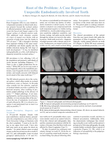
Figure 1. Intraoral radiographs and intraoral photographs
lingual surface of affected anterior teeth. sion sensitivity, palpation sensitivity, and Discussionographs
Figure 1. Intraoral radiographs and intraoral phot
A-B. Periapical radiographs demonstrating periapical pathology on 5 and 12.
This additional tooth structure consists fracture testing, and gutta percha inserted The clinical presentation of the patient
A-B. Periapical radiographs demonstrating periapical pathology on 5 and 12.
C-D. Bitewing radiographs demonstrating no carious activity and lack of periodontal
of a layer of enamel over dentin, with an through the stroma was traced to the radio- from this case report clearly falls under the
C-D. Bitewing radiographs demonstrating no carious activity and lack of periodontal
involvement on 5 and 12.
extension of pulp tissue into the dentin in lucencies. Both teeth received a diagnosis parameters of Dens Evaginatus. There is a
involvement on 5 and 12.
E-G. Intraoral photographs demonstrating clinical stomas. (E) shows an enamel
70% of cases. 1-5,7 DE is thought to develop of pulpal necrosis with a chronic apical ab- clinically visible enamel tubercle on tooth
E-G. Intraoral photographs demonstrating clinical stomas. (E) shows an enamel
tubercle on 12, and an occlusal composite restoration on 5.
from improper folding of the inner enam- scess. Furthermore, an enamel tubercle was #12 (Figure 1). While DE most commonly
tubercle on 12, and an occlusal composite restoration on 5.
el epithelium and dental papilla into the visible on #12, and a small occlusal filling presents in mandibular premolars, it is still
stellate reticulum during the bell stage of
A
A
tooth development. It may be identified B
3,7
radiographically as a thin, well-defined ra- B
diopacity toward the occlusal surface.
4
DE prevalence is low, affecting 1-4% of
the population and primarily individuals of
Asian descent (including Filipinos). 1-3,5-7
There is also a slight predilection for fe-
males. While DE can involve any tooth, it
2,7
most frequently affects the mandibular pre-
molars, least frequently involves maxillary A B
molars, and usually presents with bilateral
involvement of the same tooth type. 1-3,5-7
The DE tubercle projects above the occlu- D
C
sal surface, which often results in occlusal
C
interference, wear, or fracture. As the pulp D
often extends into the tubercle, exposure
of dentinal tubules provides a pathway for
bacterial invasion, often resulting in pre-
mature pulpal pathology in the absence of
caries. 1-3,5-7 Pulpal pathology, if not man-
aged properly, can then progress to abscess,
cellulitis, or even osteomyelitis of the jaw. C D
7
Given the risk of endodontic involvement
of teeth affected by DE, it is critical to rec- Figure 1. Intraoral radiographs and intraoral photographs
ognize it and understand how to treat it as A-B. Periapical radiographs demonstrating periapical pathology on #5 and #12.
early as possible. 2,4,6 C-D. Bitewing radiographs demonstrating no carious activity and lack of periodontal
involvement on #5 and #12.
Patient Description, Diagnosis, and E-G. Intraoral photographs demonstrating clinical stomas. (E) shows an enamel tubercle
Treatment on #12, and an occlusal composite restoration on #5.
A 28-year-old Hispanic female with
non-contributory medical history present- was visible on #5, presumably where a pre- common in premolars overall, and there is
ed to the comprehensive care clinic at New vious dentist removed an enamel tubercle. bilateral presentation. Additionally, while
York University College of Dentistry with Given this clinical and radiographic data, the characteristic radiopaque line extend-
the chief complaint, “I want to fix my fill- and the consistent presentation with the lit- ing occlusally is not visible on this patient’s
ing on my upper front tooth. I have also erature, it became clear that these teeth were periapical or bitewing radiographs, the pulp
had some spots on my gums for about six affected by Dens Evaginatus (Type V). extends farther occlusally than is typical of
months.” An initial clinical exam revealed To date, the patient has undergone end- a premolar (Figures 2A, 2B), which is an
stomas on the buccal mucosa above teeth #5 odontic therapy on tooth #5. Given the ex- indication of susceptibility to the pulpal pa-
and #12, and radiographic exam revealed tent of remaining sound tooth structure, an thology experienced.
periapical radiolucencies of #5 and #12. occlusal filling was placed following obtu-
www.nysagd.org l Fall 2019 l GP 16