
Page 200 - Libro 2
P. 200
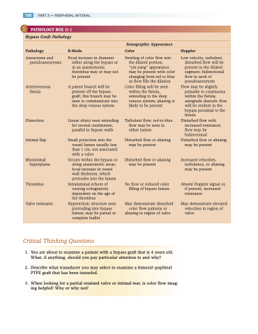
   180 PART 3 — PERIPHERAL ARTERIAL
  PATHOLOGY BOX 11-1
  Bypass Graft Pathology
 Sonographic Appearance
Pathology B-Mode Color Doppler
  Aneurysms and pseudoaneurysms
Arteriovenous fistula
Dissection
Intimal flap
Myointimal hyperplasia
Thrombus
Valve remnants
Focal increase in diameter either along the bypass or at an anastomosis; thrombus may or may not be present
A patent branch will be present off the bypass graft; this branch may be seen to communicate into the deep venous system
Linear object seen extending for several centimeters, parallel to bypass walls
Small projection into the vessel lumen usually less than 1 cm; not associated with a valve
Occurs within the bypass or along anastomotic areas; focal increase in vessel wall thickness, which protrudes into the lumen
Intraluminal echoes of varying echogenicity dependent on the age of the thrombus
Hyperechoic structure seen protruding into bypass lumen; may be partial or complete leaflet
Swirling of color flow into the dilated portion; “yin-yang” appearance may be present with color changing from red to blue as flow fills the dilation
Color filling will be seen within the fistula, extending to the deep venous system; aliasing is likely to be present
Turbulent flow; red-to-blue flow may be seen in either lumen
Low velocity, turbulent, disturbed flow will be present in the dilated segment; bidirectional flow in neck of pseudoaneurysm
Flow may be slightly pulsatile to continuous within the fistula; antegrade diastolic flow will be evident in the bypass proximal to the fistula
Disturbed flow with increased resistance; flow may be bidirectional
Disturbed flow or aliasing may be present
Increased velocities, turbulence, or aliasing may be present
Absent Doppler signal or, if present, increased resistance
May demonstrate elevated velocities in region of valve
Disturbed flow or may be present
Disturbed flow or may be present
aliasing
aliasing
No flow or reduced color filling of bypass lumen
May demonstrate disturbed color flow patterns or
aliasing in region of valve
 Critical Thinking Questions
  1. You are about to examine a patient with a bypass graft that is 4 years old. What, if anything, should you pay particular attention to and why?
2. Describe what transducer you may select to examine a femoral–popliteal PTFE graft that has been tunneled.
3. When looking for a partial retained valve or intimal tear, is color flow imag- ing helpful? Why or why not?
