
Page 199 - Libro 2
P. 199
11 — Ultrasound Assessment of Arterial Bypass Grafts
179
AB
C
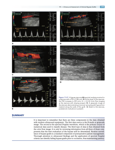
E poststenotic turbulence is present. SUMMARY
It is important to remember that there are three components to the data obtained with modern ultrasound equipment. The first data source is the B-mode or grayscale image. The second is the spectral Doppler waveform, which is the primary source of numerical data used to classify disease. The third type of data is that obtained from the color flow image. It is only by reviewing information from all three of these com- ponents that the best evaluation of the bypass will be determined. Routine surveil- lance of bypass grafts is a standard component of postoperative patient management. Thorough attention to ultrasound findings and the application of spectral Doppler criteria can identify failing bypass grafts prior to occlusion, thus maintaining patency.
D
Figure 11-21 A bypass stenosis (A) spectral analysis proximal to a stenosis with a PSV of 108 cm/s. B: At the level of the stenosis, the PSV increases to 259 cm/s, Vr 2.4. C: Color flow imaging at the stenosis with aliasing present. D: A grayscale image at the stenosis demonstrating a luminal narrowing with hypoechoic material along the walls of the graft. E: Distal to the stenosis,
