
Page 328 - Libro 2
P. 328
308
PART 5 — ABDOMINAL
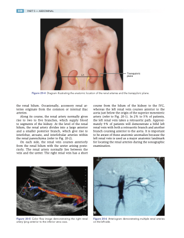
L1
Transpyloric plane
Figure 20-4 Diagram illustrating the anatomic location of the renal arteries and the transpyloric plane.
the renal hilum. Occasionally, accessory renal ar- teries originate from the common or internal iliac arteries.
Along its course, the renal artery normally gives rise to two to five branches, which supply blood to segments of the kidney. At the level of the renal hilum, the renal artery divides into a large anterior and a smaller posterior branch, which give rise to interlobar, arcuate, and interlobular arteries within the renal parenchyma (refer to Fig. 20-2).
On each side, the renal vein courses anteriorly from the renal hilum with the ureter arising poste- riorly. The renal artery normally lies between the vein and the ureter. The right renal vein has a short
Figure 20-5 Color flow image demonstrating the right renal artery lying anterior to the inferior vena cava.
course from the hilum of the kidney to the IVC, whereas the left renal vein courses anterior to the aorta just below the origin of the superior mesenteric artery (refer to Fig. 20-1). In 2% to 5% of patients, the left renal vein takes a retroaortic path. Approxi- mately 9% of patients will demonstrate a bifid left renal vein with both a retroaortic branch and another branch coursing anterior to the aorta. It is important to be aware of these anatomic anomalies because the left renal vein is used as a major anatomic landmark for locating the renal arteries during the sonographic examination.
Figure 20-6 Arteriogram demonstrating multiple renal arteries on the left side.
