
Page 330 - Libro 2
P. 330
310
PART 5 — ABDOMINAL
AB
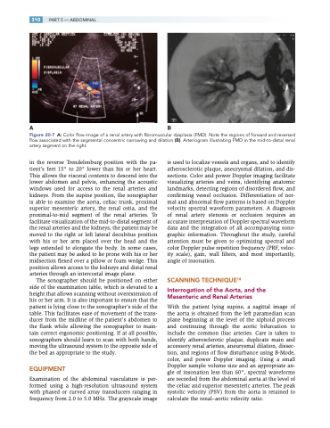
Figure 20-7 A: Color flow image of a renal artery with fibromuscular dysplasia (FMD). Note the regions of forward and reversed flow associated with the segmental concentric narrowing and dilation (B). Arteriogram illustrating FMD in the mid-to-distal renal artery segment on the right.
in the reverse Trendelenburg position with the pa- tient’s feet 15° to 20° lower than his or her heart. This allows the visceral contents to descend into the lower abdomen and pelvis, enhancing the acoustic windows used for access to the renal arteries and kidneys. From the supine position, the sonographer is able to examine the aorta, celiac trunk, proximal superior mesenteric artery, the renal ostia, and the proximal-to-mid segment of the renal arteries. To facilitate visualization of the mid-to-distal segment of the renal arteries and the kidneys, the patient may be moved to the right or left lateral decubitus position with his or her arm placed over the head and the legs extended to elongate the body. In some cases, the patient may be asked to lie prone with his or her midsection flexed over a pillow or foam wedge. This position allows access to the kidneys and distal renal arteries through an intercostal image plane.
The sonographer should be positioned on either side of the examination table, which is elevated to a height that allows scanning without overextension of his or her arm. It is also important to ensure that the patient is lying close to the sonographer’s side of the table. This facilitates ease of movement of the trans- ducer from the midline of the patient’s abdomen to the flank while allowing the sonographer to main- tain correct ergonomic positioning. If at all possible, sonographers should learn to scan with both hands, moving the ultrasound system to the opposite side of the bed as appropriate to the study.
EQUIPMENT
Examination of the abdominal vasculature is per- formed using a high-resolution ultrasound system with phased or curved array transducers ranging in frequency from 2.0 to 5.0 MHz. The grayscale image
is used to localize vessels and organs, and to identify atherosclerotic plaque, aneurysmal dilation, and dis- sections. Color and power Doppler imaging facilitate visualizing arteries and veins, identifying anatomic landmarks, detecting regions of disordered flow, and confirming vessel occlusion. Differentiation of nor- mal and abnormal flow patterns is based on Doppler velocity spectral waveform parameters. A diagnosis of renal artery stenosis or occlusion requires an accurate interpretation of Doppler spectral waveform data and the integration of all accompanying sono- graphic information. Throughout the study, careful attention must be given to optimizing spectral and color Doppler pulse repetition frequency (PRF, veloc- ity scale), gain, wall filters, and most importantly, angle of insonation.
SCANNING TECHNIQUE18
Interrogation of the Aorta, and the Mesenteric and Renal Arteries
With the patient lying supine, a sagittal image of the aorta is obtained from the left paramedian scan plane beginning at the level of the xiphoid process and continuing through the aortic bifurcation to include the common iliac arteries. Care is taken to identify atherosclerotic plaque, duplicate main and accessory renal arteries, aneurysmal dilation, dissec- tion, and regions of flow disturbance using B-Mode, color, and power Doppler imaging. Using a small Doppler sample volume size and an appropriate an- gle of insonation less than 60°, spectral waveforms are recorded from the abdominal aorta at the level of the celiac and superior mesenteric arteries. The peak systolic velocity (PSV) from the aorta is retained to calculate the renal–aortic velocity ratio.
