
Page 332 - Libro 2
P. 332
312
PART 5 — ABDOMINAL
visualize. It is helpful to image from a posterolateral plane, using the left kidney as an acoustic window. Assuming the positions of the hands on a clock, an image of the left kidney is obtained so that the renal pelvis is positioned at either 5, 6, or 7 o’clock. From the 5 o’clock position, the left renal artery will be noted to course slightly to the right before entering the kidney (Fig. 20-11). At the 6 o’clock position, the renal artery will enter the hilum on a straight course. When imaged from the 7 o’clock position, the artery will course slightly to the left just before entering the pelvis of the kidney.
Access to the distal segment of the renal artery can also be obtained by having the patient lie prone and flexed in the midsection over a pillow or foam wedge. Using an intercostal window, excellent images of the kidney and distal-to-mid segments of the renal artery are readily obtained. With both approaches, attention must be given to appropriate angle correc- tion, taking care to use a range of angles similar to those used for Doppler spectral interrogation of the proximal-to-mid segments of the artery.
Identification of Accessory Renal Arteries
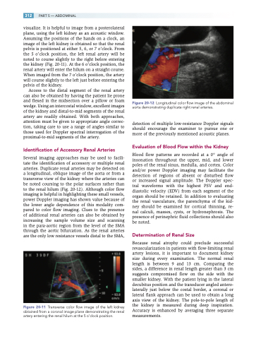
Several imaging approaches may be used to facili- tate the identification of accessory or multiple renal arteries. Duplicate renal arteries may be detected on a longitudinal, oblique image of the aorta or from a transverse view of the kidney where the arteries can be noted coursing to the polar surfaces rather than to the renal hilum (Fig. 20-12). Although color flow imaging is helpful in highlighting these small vessels, power Doppler imaging has shown value because of the lower angle dependence of this modality com- pared to color flow imaging. Clues to the presence of additional renal arteries can also be obtained by increasing the sample volume size and scanning in the para-aortic region from the level of the SMA through the aortic bifurcation. As the renal arteries are the only low resistance vessels distal to the SMA,
Figure 20-11 Transverse color flow image of the left kidney obtained from a coronal image plane demonstrating the renal artery entering the renal hilum at the 5 o’clock position.
Figure 20-12 Longitudinal color flow image of the abdominal aorta demonstrating duplicate right renal arteries.
detection of multiple low-resistance Doppler signals should encourage the examiner to pursue one or more of the previously mentioned acoustic planes.
Evaluation of Blood Flow within the Kidney
Blood flow patterns are recorded at a 0° angle of insonation throughout the upper, mid, and lower poles of the renal sinus, medulla, and cortex. Color and/or power Doppler imaging may facilitate the detection of regions of absent or disturbed flow or increased signal amplitude. The Doppler spec- tral waveforms with the highest PSV and end- diastolic velocity (EDV) from each segment of the organ should be retained. In addition to evaluating the renal vasculature, the parenchyma of the kid- ney should be examined for cortical thinning, re- nal calculi, masses, cysts, or hydronephrosis. The presence of perinephric fluid collections should also be noted.
Determination of Renal Size
Because renal atrophy could preclude successful revascularization in patients with flow-limiting renal artery lesions, it is important to document kidney size during every examination. The normal renal length is between 9 and 13 cm. Comparing the sides, a difference in renal length greater than 3 cm suggests compromised flow on the side with the smaller kidney. With the patient lying in the lateral decubitus position and the transducer angled antero- laterally just below the costal border, a coronal or lateral flank approach can be used to obtain a long axis view of the kidney. The pole-to-pole length of the kidney is measured during deep inspiration. Accuracy is enhanced by averaging three separate measurements.
