
Page 333 - Libro 2
P. 333
20 — The Renal Vasculature
313
Doppler spectral waveforms recorded within the renal parenchyma in a patient with renal vein thrombosis. Note the reversed, blunted diastolic flow component.
Evaluation of Renal Vein Thrombosis
Patients may be referred to the vascular laboratory to rule out renal vein thrombosis. Patients with acute renal failure may present with pain and hematuria. Other patients may be suspected of renal cell car- cinoma with extension of the tumor into the renal veins or IVC. The renal veins and IVC should always be examined in patients with an IVC filter or throm- bosed IVC because retrograde thrombosis may extend into the renal veins.
Confirmation of renal vein thrombosis can be technically challenging. It is important to optimize the grayscale image because acute thrombus may have acoustic properties similar to flowing blood. Although venous dilation suggests acute thrombosis, the renal vein may be contracted when the condition is chronic. The absence of an optimized spectral, color, or power Doppler signal suggests thrombosis of the renal vein. In this setting, Doppler spectral waveforms with retrograde, blunted diastolic flow
components are usually recorded throughout the arteries of the renal parenchyma (Fig. 20-13).
Use of Contrast-Enhanced Imaging
There are many factors that may influence the ability to visualize the entire length of the renal artery including excessive bowel gas, patient body habitus, and an inability to adequately position the patient to gain optimal acoustic windows.9 Quite often, imaging can be improved and diagnostic accuracy increased by employing intravenous ultrasound con- trast agents to enhance visualization of the renal arteries. Although not yet approved by the U.S. Food and Drug Administration for clinical vascular evalua- tions, investigators have shown improved diagnostic accuracy when these agents are used in the liver and in the mesenteric and peripheral vessels.17,19
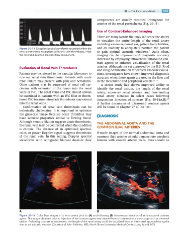
A recent study has shown improved ability to identify the renal ostium, the length of the renal artery, accessory renal arteries, and flow-limiting renal artery stenoses in select cases following intravenous injection of contrast (Fig. 20-14A,B).20 A further discussion of ultrasound contrast agents will be found in Chapter 27 of this text.
DIAGNOSIS
THE ABDOMINAL AORTA AND THE COMMON ILIAC ARTERIES
B-mode images of the normal abdominal aorta and common iliac arteries should demonstrate anechoic lumens with smooth arterial walls. Care should be
Figure 20-13
AB
Figure 20-14 Color flow images of a renal artery prior to (A) and following (B) intravenous injection of an ultrasound contrast agent. The image obtained prior to injection of the contrast agent was created from a cross-sectional aortic approach at the renal ostium. Following contrast enhancement, the length of the renal artery could be visualized from an intercostal approach using the liver as an acoustic window. (Courtesy of John Pellerito, MD, North Shore University Medical Center, Long Island, NY.)
