
Page 334 - Libro 2
P. 334
314
PART 5 — ABDOMINAL
taken to identify aneurysmal dilation, dissection, and/or atherosclerotic plaque with particular atten- tion to the regions surrounding the aortic branch vessels. Recognition of narrowed or tortuous arterial segments may be facilitated by using color flow or power Doppler imaging.
The proximal abdominal aorta carries blood to the low resistance vascular beds of the liver, spleen, and kidneys. This flow demand is reflected in the Doppler spectral waveform morphology. At this level, the Doppler spectral waveform is characterized by rapid systolic upstroke, sharp systolic peak, and forward diastolic flow. The PSV ranges from 60 to 100 cm/s. Distal to the renal arteries, the aortic waveform exhibits slightly lower velocity and a triphasic flow pattern. This waveform pattern reflects the elevated vascular resistance of the lumbar arteries and lower extremity circulation.
Flow should be laminar throughout the normal proximal abdominal aorta with the exception of slight flow disturbance, which may be evident at the ostia of the renal and mesenteric arteries. Increased velocity may be noted in tortuous vessels in the absence of disease, but high velocity, turbulent signals suggest significant arterial narrowing.
THE NORMAL RENAL ARTERY
The lumen of the normal renal artery should be an- echoic, and the vessel should have smooth walls throughout its length. Color flow and/or power Doppler imaging may demonstrate regions of nar- rowing caused by tortuosity, kinking, or extrinsic compression.
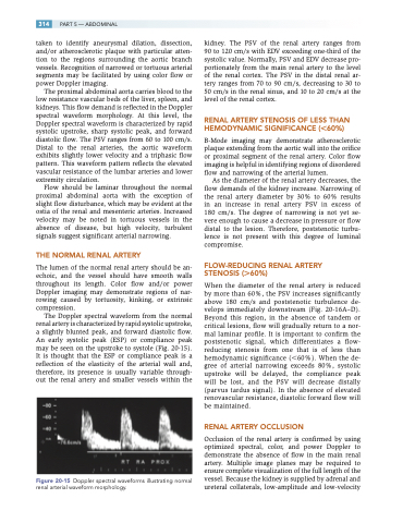
The Doppler spectral waveform from the normal renal artery is characterized by rapid systolic upstroke, a slightly blunted peak, and forward diastolic flow. An early systolic peak (ESP) or compliance peak may be seen on the upstroke to systole (Fig. 20-15). It is thought that the ESP or compliance peak is a reflection of the elasticity of the arterial wall and, therefore, its presence is usually variable through- out the renal artery and smaller vessels within the
Figure 20-15 Doppler spectral waveforms illustrating normal renal arterial waveform morphology.
kidney. The PSV of the renal artery ranges from 90 to 120 cm/s with EDV exceeding one-third of the systolic value. Normally, PSV and EDV decrease pro- portionately from the main renal artery to the level of the renal cortex. The PSV in the distal renal ar- tery ranges from 70 to 90 cm/s, decreasing to 30 to 50 cm/s in the renal sinus, and 10 to 20 cm/s at the level of the renal cortex.
RENAL ARTERY STENOSIS OF LESS THAN HEMODYNAMIC SIGNIFICANCE (60%)
B-Mode imaging may demonstrate atherosclerotic plaque extending from the aortic wall into the orifice or proximal segment of the renal artery. Color flow imaging is helpful in identifying regions of disordered flow and narrowing of the arterial lumen.
As the diameter of the renal artery decreases, the flow demands of the kidney increase. Narrowing of the renal artery diameter by 30% to 60% results in an increase in renal artery PSV in excess of 180 cm/s. The degree of narrowing is not yet se- vere enough to cause a decrease in pressure or flow distal to the lesion. Therefore, poststenotic turbu- lence is not present with this degree of luminal compromise.
FLOW-REDUCING RENAL ARTERY STENOSIS (60%)
When the diameter of the renal artery is reduced by more than 60%, the PSV increases significantly above 180 cm/s and poststenotic turbulence de- velops immediately downstream (Fig. 20-16A–D). Beyond this region, in the absence of tandem or critical lesions, flow will gradually return to a nor- mal laminar profile. It is important to confirm the poststenotic signal, which differentiates a flow- reducing stenosis from one that is of less than hemodynamic significance (60%). When the de- gree of arterial narrowing exceeds 80%, systolic upstroke will be delayed, the compliance peak will be lost, and the PSV will decrease distally (parvus tardus signal). In the absence of elevated renovascular resistance, diastolic forward flow will be maintained.
RENAL ARTERY OCCLUSION
Occlusion of the renal artery is confirmed by using optimized spectral, color, and power Doppler to demonstrate the absence of flow in the main renal artery. Multiple image planes may be required to ensure complete visualization of the full length of the vessel. Because the kidney is supplied by adrenal and ureteral collaterals, low-amplitude and low-velocity
