
Page 329 - Libro 2
P. 329
ETIOLOGY OF RENAL ARTERY DISEASE
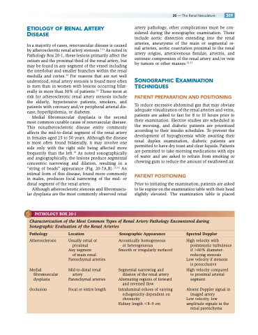
In a majority of cases, renovascular disease is caused by atherosclerotic renal artery stenosis.7,11 As noted in Pathology Box 20-1, these lesions primarily affect the ostium and the proximal third of the renal artery, but may be found in any segment of the vessel including the interlobar and smaller branches within the renal medulla and cortex.12 For reasons that are not well understood, renal artery stenosis is found more often in men than in women with lesions occurring bilat- erally in more than 30% of patients.13 Those most at risk for atherosclerotic renal artery stenosis include the elderly, hypertensive patients, smokers, and patients with coronary and/or peripheral arterial dis- ease, hyperlipidemia, or diabetes.
Medial fibromuscular dysplasia is the second most common curable cause of renovascular disease. This nonatherosclerotic disease entity commonly affects the mid-to-distal segment of the renal artery in females aged 25 to 50 years. Although the disease is most often found bilaterally, it may involve one side only with the right side being affected more frequently than the left.14 As noted sonographically and angiographically, the lesions produce segmental concentric narrowing and dilation, resulting in a “string of beads” appearance (Fig. 20-7A,B).15,16 An intimal form of this disease, found more commonly in males, produces focal narrowing of the mid- or distal segment of the renal artery.
Although atherosclerotic stenosis and fibromuscu- lar dysplasia are the most commonly observed renal
artery pathology, other complications must be con- sidered during the sonographic examination. These include aortic dissection extending into the renal arteries, aneurysms of the main or segmental re- nal arteries, aortic coarctation proximal to the renal artery origins, arteriovenous fistulae, arteritis, and extrinsic compression of the renal artery and/or vein by tumors or other masses.12,17
SONOGRAPHIC EXAMINATION TECHNIQUES
PATIENT PREPARATION AND POSITIONING
To reduce excessive abdominal gas that may obviate adequate visualization of the renal arteries and veins, patients are asked to fast for 8 to 10 hours prior to their examination. Elective studies are scheduled in the morning, and diabetic patients are prioritized according to their insulin schedules. To prevent the development of hypoglycemia while awaiting their renal duplex examination, diabetic patients are permitted to have dry toast and clear liquids. Patients are permitted to take morning medications with sips of water and are asked to refrain from smoking or chewing gum to reduce the amount of swallowed air.
PATIENT POSITIONING
Prior to initiating the examination, patients are asked to lie supine on the examination table with their head slightly elevated. The examination table is placed
20 — The Renal Vasculature 309
PATHOLOGY BOX 20-1
Characterization of the Most Common Types of Renal Artery Pathology Encountered during Sonographic Evaluation of the Renal Arteries
Pathology Location Sonographic Appearance Spectral Doppler
Atherosclerosis
Medial fibromuscular dysplasia
Occlusion
Usually ostial or proximal
Any segment
of main renal
Parenchymal arteries
Mid-to-distal renal artery
Parenchymal arteries Focal or entire length
Acoustically homogeneous or heterogeneous
Smooth or irregularly surfaced
Segmental narrowing and dilation of the renal artery
Alternating regions of forward and reversed flow
Intraluminal echoes of varying echogenicity dependent on chronicity
Kidney length 8–9 cm
High velocity with poststenotic turbulence if 60% diameter- reducing stenosis
Low velocity if stenosis is preocclusive
High velocity compared to proximal arterial segment
Absent Doppler signal in imaged artery
Low velocity, low amplitude signals in the
renal parenchyma
