
Page 371 - Libro 2
P. 371
23 — Evaluation of Kidney and Liver Transplants
351
renal vein is typically anastomosed to the recipient external iliac vein in an end-to-side approach. The ureteral anastomosis is most commonly made by creating an ureteroneocystostomy—implanting the donor ureter into the dome of the bladder above the native ureteral orifice (UVJ).
Rarely, two pediatric donor kidneys may be trans- planted en bloc into an adult recipient. In such cases, the donor aorta with the main renal arteries attached is harvested along with the two kidneys, and a direct end-to-side anastomosis between the donor aorta and the recipient external artery is created. Similarly, the donor inferior vena cava (IVC) receiving both the right and left main renal veins is implanted end to side with the recipient iliac vein.
Although practice varies nationwide, placement of an external drain next to the kidney is believed to decrease the incidence of lymphoceles formation, a relatively common postsurgical complication that can cause graft dysfunction by compressing the re- nal parenchyma or vascular/ureteral anastomoses. Superinfection may also occur. Ureteral stents from the intrarenal collecting system into the bladder are commonly placed to reduce the likelihood of ureteral scarring or necrosis as well as extravasation of urine, which may lead to the development of urinomas. In most patients, the native kidneys are left in place.
SONOGRAPHIC EXAMINATION TECHNIQUES
PATIENT PREPARATION
Typically, no patient preparation is needed to eval- uate a renal transplant. It may be helpful for the patient to have some urine in the bladder. It is impor- tant for the sonographer to review the surgical notes or speak to the surgeon before the initial or base- line ultrasound is obtained. The sonographer should know the following information at a minimum: loca- tion of the kidney, if a single kidney or two pediatric kidneys were placed, which native vessels were used to anastomose with the main renal artery and vein, any vascular anomalies such as duplicated vessels, and any additional information that may affect the ultrasound examination. The sonographer should also review any recent studies, especially if pathol- ogy was present as well as to review the vascular anatomy and anastomotic sites.
PATIENT POSITIONING
The patient is examined in the supine position. If bowel or gas is obscuring part of the kidney, the patient may be turned into an oblique or decubitus
position to try to improve visualization of the kidney.
EQUIPMENT
A 3- to 5-MHz curved linear array transducer can be used. This will provide an aperture to allow for insonation of the entire kidney. The transducer can be gently rocked to push bowel gas out of the way to improve visualization of the kidney. Varying the imaging depth will be necessary due to the superfi- cial placement of most transplanted kidneys. In thin patients, a higher transducer frequency or harmonic imaging may improve resolution.
SCANNING TECHNIQUE
Imaging protocols must be based on current accredi- tation guidelines and should be reviewed annually. These guidelines can be found at http://www.icavl. org, http://www.aium.org, and http://www.acr.org.
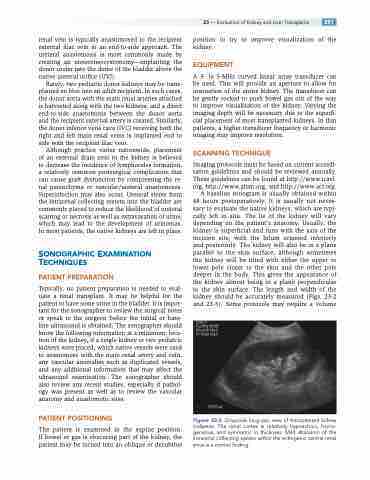
A baseline sonogram is usually obtained within 48 hours postoperatively. It is usually not neces- sary to evaluate the native kidneys, which are typi- cally left in situ. The lie of the kidney will vary depending on the patient’s anatomy. Usually, the kidney is superficial and runs with the axis of the incision site, with the hilum oriented inferiorly and posteriorly. The kidney will also be in a plane parallel to the skin surface, although sometimes the kidney will be tilted with either the upper or lower pole closer to the skin and the other pole deeper in the body. This gives the appearance of the kidney almost being in a plane perpendicular to the skin surface. The length and width of the kidney should be accurately measured (Figs. 23-2 and 23-3). Some protocols may require a volume
Figure 23-2 Grayscale long-axis view of transplanted kidney (calipers). The renal cortex is relatively hypoechoic, homo- geneous, and symmetric in thickness. Mild dilatation of the intrarenal collecting system within the echogenic central renal sinus is a normal finding.
