
Page 372 - Libro 2
P. 372
352
PART 5 — ABDOMINAL
Figure 23-3 Grayscale transverse view of a transplanted kidney.
measurement of the kidney, which would require measuring the kidney in all three planes: length, width, and anterioposterior (AP) dimensions. The transplanted kidney will look like a normal kid- ney in shape and echotexture. A comparison view with the liver or spleen cannot be obtained as the kidney is now located in the pelvis.
Grayscale
Grayscale imaging should be performed first. Doc- umented images should include views of the long axis of the kidney in the mid-axis to measure the length of the kidney as well as longitudinal views of the lateral and medial aspects of the kidney. Re- nal length is variable but is often slightly larger than the native kidney as the kidney will hypertro- phy, usually reaching its maximal size by 6 months postop. Increase or decrease in renal length from one exam to the next is a nonspecific indicator of graft dysfunction. Next, views that are 90° to the long axis of the kidney (i.e., in the transverse
plane) are obtained. Many laboratories obtain transverse views superior to the kidney, through the upper pole, mid-pole with transverse and AP measurements, lower pole, and finally, inferior to the kidney. The superior and inferior transverse views are used to evaluate for any perinephric fluid collections. However, a general survey should also be performed to look for fluid collections. Multiple longitudinal and transverse images of the blad- der, or the bladder area if the patient has a Foley catheter in place, should be obtained. An oblique view showing the lower pole of the kidney and the bladder in the same image can also be recorded. This is a helpful image to evaluate for the presence of a urinoma. Any fluid collection seen near the bladder will require further investigation either by having the patient void or by instilling appropriate fluid through the patient’s catheter to distend the bladder.
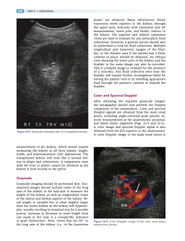
Color and Spectral Doppler
After obtaining the requisite grayscale images, the sonographer should now perform the Doppler component of the examination. Color and spectral Doppler signals are obtained from the main renal artery, including angle-corrected peak systolic ve- locity measurements at the anastomosis, proximal, and distal (hilar) segments (Figs. 23-4 and 23-5). A color image and spectral Doppler signal is also obtained from the EIA superior to the anastomosis. A color Doppler image of the main renal artery is
Figure 23-4 Color Doppler image of the main renal artery anastomosis (arrow).
