
Page 173 - Libro vascular I
P. 173
Chap-12.qxd 29~8~04 14:52 Page 164
164
PERIPHERAL VASCULAR ULTRASOUND
INTRODUCTION
Venous disorders are a common problem and con- sume a significant proportion of the resources avail- able to health care systems. Approximately 20–25% of women and 10–15% of men have visible varicose veins (Callam 1994). Significant venous disease can lead to venous ulceration, resulting in a marked loss in quality of life. Duplex scanning has had a dra- matic impact on the noninvasive assessment of the venous system, and it is now the most commonly performed procedure for the detailed investigation of lower limb venous insufficiency. Lower limb venous duplex imaging can be used for the assess- ment of patients with primary or secondary varicose veins or for the investigation of patients with skin changes and venous ulceration. In comparison with arterial duplex scanning, venous duplex investiga- tions can be technically challenging due to the wide range of anatomical variations in the venous system. This chapter covers the basic anatomy of the venous system and scanning techniques used for the assess- ment of lower limb venous insufficiency.
ANATOMY
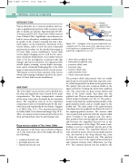
The lower limb venous system can be divided into the deep and superficial veins, located in two main compartments. The deep compartment contains all the deep veins and is bounded by the muscular fascia. The superficial veins lie in the superficial compartment and are bounded deeply by the mus- cular fascia and superficially by the dermis (Caggiati et al 2002) (Fig. 12.1). The muscular fascial layer is usually visible on an ultrasound image (Fig. 12.2). There are numerous interconnections between the deep and superficial veins via perforating veins.
Deep venous system of the lower limbs
The anatomy of the deep veins is shown in Figure 12.3. The main deep veins of the thigh and calf are the following:
● common femoral vein
● profunda femoris vein
● superficial femoral vein
SC
Dermis Superficial branch of LSV
LSV Muscular
DP fascia Deep vein
Perforator
A diagram of the deep and superficial vein compartments. The main trunk of the saphenous vein lies
in the saphenous compartment (SC), located within the superficial compartment; see text.
Figure 12.1
● above-knee popliteal vein
● below-knee popliteal vein
● posterior tibial veins
● peroneal veins
● anterior tibial veins
● gastrocnemius veins
● soleal veins and sinuses.
The posterior tibial and peroneal veins are usually paired and are associated with their respective arter- ies, which frequently lie in between the paired veins. The paired veins join into common trunks in the upper calf before forming the below-knee popliteal vein. The soleal veins are deep venous sinuses and veins of the soleus muscle that drain into the popliteal vein. They are an important part of the calf muscle pump mechanism (see Ch. 5). The gastroc- nemius veins drain the medial and lateral sides of the gastrocnemius muscle and are usually larger in the medial side. The main trunk of the gastrocnemius vein drains into the popliteal vein below the level of the saphenopopliteal junction. The anterior tibial vein is paired and associated with the anterior tibial artery. It drains to the popliteal vein. The above- knee popliteal vein runs through the adductor canal and becomes the superficial femoral vein in the lower medial aspect of the thigh. The name is mis- leading as it is not a superficial vein but part of the deep venous system. The superficial femoral vein runs toward the groin, where the profunda femoris vein, also known as the deep femoral vein, joins to form the common femoral vein. This junction lies below the level of the saphenofemoral junction and common femoral artery bifurcation (see Fig. 9.6). The common femoral vein lies medial to the
