
Page 174 - Libro vascular I
P. 174
Chap-12.qxd 29~8~04 14:52 Page 165
ANATOMY OF THE LOWER LIMB VENOUS SYSTEM AND ASSESSMENT OF VENOUS INSUFFICIENCY
V
Right renal vein Inferior vena cava
Right common iliac vein
Right external iliac vein
Right common femoral vein Saphenofemoral junction Profunda femoris vein (deep femoral vein)
Right superficial femoral vein
Saphenopopliteal junction Gastrocnemius vein system Popliteal vein
Soleal veins and sinuses
Anterior tibial veins Peroneal veins
Posterior tibial veins
A
LG
B
V
MG
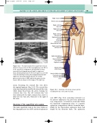
Figure 12.2
artery, becoming the external iliac vein above the inguinal ligament (Fig. 12.3). The external iliac vein runs deep and is joined by the internal iliac vein, which drains blood from the pelvis, forming the common iliac vein. The left common iliac vein runs underneath the right common iliac artery to drain into the vena cava, which lies to the right of the aorta.
Anatomy of the superficial vein system
The main superficial veins in the lower limbs are the long saphenous vein (LSV) and short saphenous
The main trunks of the superficial veins are shown in cross section. A: The long saphenous vein (V) lies in the superficial compartment, bounded by the deep muscular fascia (upward arrows) and the saphenous fascia (downward arrows). B: The short saphenous vein (V) is also bounded by the deep fascia (upward arrows) and saphenous fascia (downward arrows). The medial gastrocnemius muscle (MG) and lateral gastrocnemius muscle (LG) are shown on this image of the right leg.
Anatomy of the deep venous system, including the iliac veins and vena cava.
Figure 12.3
vein (SSV) (Fig. 12.4) (sometimes referred to as the greater saphenous vein and lesser saphenous vein, respectively). It should be noted that within the superficial compartment there is a separate saphenous compartment which is bounded super- ficially by the hyperechoic saphenous fascia and deeply by the muscular fascia. The saphenous
165
