
Page 20 - Libro vascular I
P. 20
Chap-02.qxd 29~8~04 13:19 Page 11
ULTRASOUND AND IMAGING
11
the frequency (i.e., f 4) for structures that are much smaller than the wavelength of the ultrasound]. In the case of peripheral vascular ultrasound, specular reflection will occur at the vessel walls, which are often perpendicular to the beam, leading to large reflected signals. However, ultrasound will be scat- tered by groups of red blood cells within the lumen, leading to much smaller returning signals, which will not normally be visible on an image.
LOSS OF ULTRASOUND ENERGY
IN TISSUE
Attenuation is the loss of energy from the ultra- sound beam as it passes through tissue. The more the ultrasound energy is attenuated by the tissue, the less energy will be available to return to the transducer or to penetrate deeper into the tissue. Attenuation is caused by several different processes. These include absorption, scattering, reflection and beam divergence. Absorption causes ultrasound energy to be converted into heat as the beam passes through the tissue. The rate of absorption varies in different types of tissue. Ultrasound energy can also be lost by scattering from small structures within the tissue or reflection from large bound- aries that are not perpendicular to the beam, pre- venting the ultrasound from returning to the transducer. The attenuation coefficients of various tissues are presented in Table 2.3, from which it can be seen that muscle attenuates the ultrasound more quickly than fat. The units of the coefficient of attenuation are in dBMHz1cm1, showing that the rate of attenuation depends on frequency of ultrasound, with higher frequencies being
in vascular ultrasound, with the exception of the presence of the skull bone in the path of a tran- scranial Doppler beam.
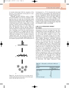
Although specular reflection occurs at large, smooth boundaries, the majority of signals return- ing from tissue are made up of ultrasound energy that has been back-scattered from rough surfaces or small structures within the tissue. When the ultra- sound beam interacts with a rough surface or small structure it will be scattered in all directions rather than reflected back along one path. Figure 2.8 shows the difference between specular reflection and scattering from rough surfaces and small struc- tures. Scattering occurs when the small structures are of a similar size to or smaller than the wave- length of the ultrasound and will result in less of the ultrasound returning to the transducer along the original beam path. The amount of energy lost from the beam by scattering is highly dependent on the frequency [proportional to the fourth power of
A
B
Table 2.3 Attenuation coefficients of different tissues
Medium
Attenuation coefficient at 1 MHz (dB cm1)
Water (20° C) 0.2 Fat 60 Blood 20 Muscle 150 Bone 1000 Soft tissue (average) 70
C
Figure 2.8 Specular reflections occur at large smooth interfaces (A), whereas ultrasound is scattered by rough surfaces (B) and small structures (C).
