
Page 39 - Libro vascular I
P. 39
Chap-03.qxd 29~8~04 13:20 Page 30
30
PERIPHERAL VASCULAR ULTRASOUND
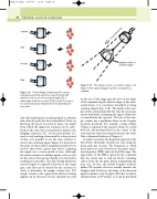
Transmit
A
Wait
Receive
Wait
and then only receives from a given depth (C); it
waits again, with the receiver off (D), for all the echoes to return from greater depths before transmitting the next pulse.
and only acquiring the returning signal at a known time after the pulse has been transmitted. Thus, by knowing the speed of sound in tissue, the depth from which the signal has returned can be calcu- lated, in the same way as described for pulsed echo imaging (equation 2.3). As the piezoelectric ele- ment is only emitting ultrasound for a short period of time, it is possible to use the same element to receive the returning signal. Figure 3.9 shows how the pulse of ultrasound is transmitted and how the receiver then waits a given time before acquiring the signal over a short period of time. Although the system acquires no further signals, it has to wait for the echoes from greater depths to return before sending the next pulse. The time during which the received signal is acquired is known as the range gate, and this can be altered by the operator in order to determine the sample volume size. The sample volume is the region from which returning signals can be detected. Its size depends not only
Figure 3.9
B
C
D Figure 3.10
single element pulsed Doppler system is shaped like a
Sample volume, or sensitive region
Pulsed Doppler ultrasound. The system transmits a pulse (A), waits for a specified time (B)
teardrop.
The sample volume, or sensitive region, of a
on the size of the range gate but also on the shape of the transmitted pulse and the shape of the ultra- sound beam; it is sometimes described as being teardrop-shaped (Fig. 3.10). The depth of the sam- ple volume is determined by the time the electronic circuit waits before acquiring the signal, which also is controlled by the operator. The size of the sam- ple volume has a significant effect on the Doppler spectrum produced. For example, a large sample volume is required if the operator wishes to record both the fast-moving blood in the center of the vessel and the slower moving blood near the walls. This is discussed further in Chapter 6.
In order to measure the frequencies present in the blood flow, thousands of pulses are sent along the beam path per second. The frequency at which these pulses are sent is known as the pulse repeti- tion frequency (PRF) and is in the kHz range. The upper limit of the PRF is given by the constraint that the system has to wait for all the returning echoes from the last pulse before transmitting the next one. In fact, the pulsed Doppler method, unlike CW systems, does not actually measure the Doppler shift. However, the shape of the detected signal is similar to the Doppler shift that would be obtained from a CW system, so it can be described
Transmitted pulse
