
Page 66 - Libro vascular I
P. 66
Chap-05.qxd 29~8~04 13:25 Page 57
BLOOD FLOW AND ITS APPEARANCE ON COLOR FLOW IMAGING
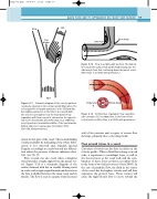
ECA
ICA
Flow divider
Flow divider
Inner wall
Flow in a right-angle junction. The dashed line shows the surface that divides fluid flowing into the
side branch from that continuing down the parent vessel. (After Caro et al 1978, with permission.)
Figure 5.14
CCA
A
Inside bend
Figure 5.13
Outside bend
helical vortices. (After Caro et al 1978, with permission.)
Schematic diagram of the velocity patterns
commonly observed in the normal carotid bifurcation. The B velocity profile is flat and symmetric in the CCA and flat
but slightly asymmetric in the ICA. In the carotid bulb
the velocities are highest near the flow divider. Flow
separation with flow reversal is observed on the opposite
side to the flow divider. (From Reneman et al 1985 Flow
velocity patterns in and distensibility of the carotid artery
bulb in subjects of various ages. Circulation 71(3):
500–509, with permission.)
Figure 5.15
A: Distortion of parabolic flow caused by tube curvature. B: Secondary flow, in the form of two
57
wall of the junction and a region of reverse flow develops, primarily due to the sharp bend.
Flow around curves in a vessel
Curvature of vessels can also have an effect on the velocity profile. When a fluid flows along a curved tube, it experiences a centrifugal force, as well as the viscous forces at the vessel wall, and the com- bination of these forces results in secondary flow, in the form of two helical vortices (Oates 2003). In the case of parabolic flow, the fluid in the center of the vessel has the highest velocity and will thus experience the greatest force. These vortices will cause the high-velocity flow to move toward the
systole in that part of the vessel. This normal finding could potentially be misleading if the whole bifur- c a t i o n i s n o t o b s e r v e d a n d , t y p i c a l l y, s p e c t r a l Doppler recordings are made beyond the bifurca- tion unless the presence of disease indicates other- wise (see Ch. 8).
Flow reversal can also occur when a daughter vessel branches at right angles from the parent ves- sel. Figure 5.14 is a schematic diagram of the results obtained with dye in steadily flowing water in a tube with a right-angled branch and shows how the flow is divided between the main vessel and its branch. The flow is seen to separate from the inner
