
Page 127 - Basic _ Clinical Pharmacology ( PDFDrive )
P. 127
CHAPTER 7 Cholinoceptor-Activating & Cholinesterase-Inhibiting Drugs 113
sympathetic-parasympathetic interaction is complex because Parasympathetic nerves can regulate arteriolar tone in vascular
muscarinic modulation of sympathetic influences occurs by beds in thoracic and abdominal visceral organs. Acetylcholine
inhibition of norepinephrine release and by postjunctional released from postganglionic parasympathetic nerves relaxes
cellular effects. Muscarinic receptors that are present on post- coronary arteriolar smooth muscle via the NO/cGMP pathway
ganglionic parasympathetic nerve terminals allow neurally in humans as described above. Damage to the endothelium, as
released acetylcholine to inhibit its own secretion. The neuronal occurs with atherosclerosis, eliminates this action, and acetylcho-
muscarinic receptors need not be the same subtype as found on line is then able to contract arterial smooth muscle and produce
effector cells. Therefore, the net effect on heart rate depends on vasoconstriction. Parasympathetic nerve stimulation also causes
local concentrations of the agonist in the heart and in the vessels vasodilation in cerebral blood vessels; however, the effect often
and on the level of reflex responsiveness. appears as a result of NO released either from NANC (nitrergic)
Parasympathetic innervation of the ventricles is much less neurons or as a cotransmitter from cholinergic nerves. The relative
extensive than that of the atria; activation of ventricular musca- contributions of cholinergic and NANC neurons to the vascular
rinic receptors causes much less direct physiologic effect than that effects of parasympathetic nerve stimulation are not known for
seen in atria. However, the indirect effects of muscarinic agonists most viscera. Skeletal muscle receives sympathetic cholinergic
on ventricular function are clearly evident during sympathetic vasodilator nerves, but the view that acetylcholine causes vasodi-
nerve stimulation because of muscarinic modulation of sympa- lation in this vascular bed has not been verified experimentally.
thetic effects (“accentuated antagonism”). Nitric oxide, rather than acetylcholine, may be released from these
In the intact organism, intravascular injection of muscarinic neurons. However, this vascular bed responds to exogenous cho-
agonists produces marked vasodilation. However, earlier studies line esters because of the presence of M receptors on endothelial
3
of isolated blood vessels often showed a contractile response to and smooth muscle cells.
these agents. It is now known that acetylcholine-induced vaso- The cardiovascular effects of all the choline esters are similar
dilation arises from activation of M receptors and requires the to those of acetylcholine—the main difference being in their
3
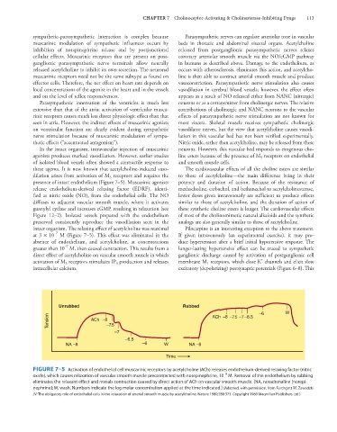
presence of intact endothelium (Figure 7–5). Muscarinic agonists potency and duration of action. Because of the resistance of
release endothelium-derived relaxing factor (EDRF), identi- methacholine, carbachol, and bethanechol to acetylcholinesterase,
fied as nitric oxide (NO), from the endothelial cells. The NO lower doses given intravenously are sufficient to produce effects
diffuses to adjacent vascular smooth muscle, where it activates similar to those of acetylcholine, and the duration of action of
guanylyl cyclase and increases cGMP, resulting in relaxation (see these synthetic choline esters is longer. The cardiovascular effects
Figure 12–2). Isolated vessels prepared with the endothelium of most of the cholinomimetic natural alkaloids and the synthetic
preserved consistently reproduce the vasodilation seen in the analogs are also generally similar to those of acetylcholine.
intact organism. The relaxing effect of acetylcholine was maximal Pilocarpine is an interesting exception to the above statement.
−7
at 3 × 10 M (Figure 7–5). This effect was eliminated in the If given intravenously (an experimental exercise), it may pro-
absence of endothelium, and acetylcholine, at concentrations duce hypertension after a brief initial hypotensive response. The
−7
greater than 10 M, then caused contraction. This results from a longer-lasting hypertensive effect can be traced to sympathetic
direct effect of acetylcholine on vascular smooth muscle in which ganglionic discharge caused by activation of postganglionic cell
+
activation of M receptors stimulates IP production and releases membrane M receptors, which close K channels and elicit slow
3
1
3
intracellular calcium. excitatory (depolarizing) postsynaptic potentials (Figure 6–8). This
Unrubbed Rubbed
–6 W
Tension ACh –8 –7.5
ACh
–8 –7.5 –7–6.5
–7
–6.5
NA –8 –6 W NA –8
Time
FIGURE 7–5 Activation of endothelial cell muscarinic receptors by acetylcholine (ACh) releases endothelium-derived relaxing factor (nitric
−8
oxide), which causes relaxation of vascular smooth muscle precontracted with norepinephrine, 10 M. Removal of the endothelium by rubbing
eliminates the relaxant effect and reveals contraction caused by direct action of ACh on vascular smooth muscle. (NA, noradrenaline [norepi-
nephrine]; W, wash. Numbers indicate the log molar concentration applied at the time indicated.) (Adapted, with permission, from Furchgott RF, Zawadzki
JV: The obligatory role of endothelial cells in the relaxation of arterial smooth muscle by acetylcholine. Nature 1980;288:373. Copyright 1980 Macmillan Publishers Ltd.)