
Page 122 - Basic _ Clinical Pharmacology ( PDFDrive )
P. 122
108 SECTION II Autonomic Drugs
Cholinoceptor stimulants
Heart and Glands and
Nerve smooth muscle endothelium
Alkaloids Reversible
Muscarinic
Direct-acting Receptors ACh Indirect-acting
drugs Nicotinic drugs
Choline esters Irreversible
Neuromuscular Autonomic Central
end plate, ganglion nervous
skeletal muscle cells system
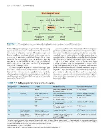
FIGURE 7–1 The major groups of cholinoceptor-activating drugs, receptors, and target tissues. ACh, acetylcholine.
Conceivably, agonist or antagonist ligands could signal by chang- Nonselective cholinoceptor stimulants in sufficient dosage can
ing the quaternary structure of the receptor, that is, the ratio of produce very diffuse and marked alterations in organ system func-
monomeric to oligomeric receptors. Muscarinic receptors are tion because acetylcholine has multiple sites of action where it ini-
located on plasma membranes of cells in the central nervous tiates both excitatory and inhibitory effects. Fortunately, drugs are
system and in autonomic ganglia (see Figure 6–8), in organs available that have a degree of selectivity, so that desired effects can
innervated by parasympathetic nerves as well as on some tis- often be achieved while avoiding or minimizing adverse effects.
sues that are not innervated by these nerves, eg, endothelial cells Selectivity of action is based on several factors. Some drugs
(Table 7–1), and on those tissues innervated by postganglionic stimulate either muscarinic receptors or nicotinic receptors selec-
sympathetic cholinergic nerves. tively. Some agents stimulate nicotinic receptors at neuromuscular
Nicotinic receptors are part of a transmembrane polypep- junctions preferentially and have less effect on nicotinic receptors
tide whose subunits form cation-selective ion channels (see in ganglia. Organ selectivity can also be achieved by using appro-
Figure 2–9). These receptors are located on plasma membranes priate routes of administration (“pharmacokinetic selectivity”).
of postganglionic cells in all autonomic ganglia, of muscles inner- For example, muscarinic stimulants can be administered topically
vated by somatic motor fibers, and of some central nervous system to the surface of the eye to modify ocular function while minimiz-
neurons (see Figure 6–1). ing systemic effects.
TABLE 7–1 Subtypes and characteristics of cholinoceptors.
Receptor Type Other Names Location Structural Features Postreceptor Mechanism
Nerves Seven transmembrane segments, IP 3 , DAG cascade
M 1
G q/11 protein-linked
M 2 Cardiac M 2 Heart, nerves, smooth Seven transmembrane segments, Inhibition of cAMP production,
+
muscle G i/o protein-linked activation of K channels
Glands, smooth muscle, Seven transmembrane segments, IP 3 , DAG cascade
M 3
endothelium G q/11 protein-linked
M 4 CNS Seven transmembrane segments, Inhibition of cAMP production
G i/o protein-linked
CNS Seven transmembrane segments, IP 3 , DAG cascade
M 5
G q/11 protein-linked
+
+
1
N M Muscle type, end Skeletal muscle Pentamer [(α1) 2 β1δγ)] Na , K depolarizing ion channel
plate receptor neuromuscular junction
+
+
1
Neuronal type, CNS, postganglionic cell Pentamer with α and β subunits Na , K depolarizing ion channel
N N
ganglion receptor body, dendrites only, eg, (α4) 2 (β2) 3 (CNS) or
α3α5(β2) 3 (ganglia)
1
Pentameric structure in Torpedo electric organ and fetal mammalian muscle has two α1 subunits and one each of β1, δ, and γ subunits. The stoichiometry is indicated by
subscripts, eg, [(α1) 2 β1 δ γ]. In adult muscle, the γ subunit is replaced by an ε subunit. There are 12 neuronal nicotinic receptors with nine α (α2-α10) subunits and three
(β2-β4) subunits. The subunit composition varies among different mammalian tissues.
DAG, diacylglycerol; IP 3 , inositol trisphosphate.
Data from Millar NS, Gotti C: Diversity of vertebrate nicotinic receptors. Neuropharmacology 2009;56:237.