
Page 117 - Basic _ Clinical Pharmacology ( PDFDrive )
P. 117
CHAPTER 6 Introduction to Autonomic Pharmacology 103
Preganglionic Membrane
axon potential
0 Spike
Slow Late, slow
EPSP IPSP EPSP EPSP
mV
Peptides
M 1
Electrode N N
M 2 (Receptor types)
–100
Postganglionic Milliseconds Seconds Minutes
neuron
Time
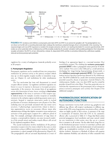
FIGURE 6–8 Excitatory and inhibitory postsynaptic potentials (EPSP and IPSP) in an autonomic ganglion cell. The postganglionic neu-
ron shown at the left with a recording electrode might undergo the membrane potential changes shown schematically in the recording. The
response begins with two EPSP responses to nicotinic (N) receptor activation, the first not reaching threshold. The second, suprathreshold, EPSP
evokes an action potential, which is followed by an IPSP, probably evoked by M 2 receptor activation (with possible participation from dopamine
receptor activation). The IPSP is, in turn, followed by a slower, M 1 -dependent EPSP, and this is sometimes followed by a still slower peptide-
induced excitatory postsynaptic potential.
regulation by a variety of endogenous chemicals probably occurs binding of an appropriate ligand to a neuronal nicotinic (N )
N
at all synapses. acetylcholine receptor. The resulting fast excitatory postsynaptic
potential (EPSP) evokes a propagated action potential if thresh-
C. Postsynaptic Regulation old is reached. This event is often followed by a small and slowly
Postsynaptic regulation can be considered from two perspectives: developing but longer-lasting hyperpolarizing afterpotential—a
modulation by previous activity at the primary receptor (which slow inhibitory postsynaptic potential (IPSP). This hyperpolar-
may up- or down-regulate receptor number or desensitize recep- ization involves opening of potassium channels by M cholinocep-
2
tors; see Chapter 2), and modulation by other simultaneous tors. The IPSP is followed by a small, slow excitatory postsynaptic
events. potential caused by closure of potassium channels linked to M
1
The first mechanism has been well documented in several cholinoceptors. Finally, a late, very slow EPSP may be evoked by
receptor-effector systems. Up-regulation and down-regulation are peptides released from other fibers. These slow potentials serve to
known to occur in response to decreased or increased activation, modulate the responsiveness of the postsynaptic cell to subsequent
respectively, of the receptors. An extreme form of up-regulation primary excitatory presynaptic nerve activity. (See Chapter 21 for
occurs after denervation of some tissues, resulting in denervation additional examples.)
supersensitivity of the tissue to activators of that receptor type.
In skeletal muscle, for example, nicotinic receptors are normally
restricted to the end plate regions underlying somatic motor nerve PHARMACOLOGIC MODIFICATION OF
terminals. Surgical or traumatic denervation results in marked AUTONOMIC FUNCTION
proliferation of nicotinic cholinoceptors over all parts of the fiber,
including areas not previously associated with any motor nerve Because transmission involves both common (eg, ganglionic) and
junctions. A pharmacologic supersensitivity related to denerva- different (eg, effector cell receptor) mechanisms in different seg-
tion supersensitivity occurs in autonomic effector tissues after ments of the ANS, some drugs produce less selective effects, whereas
administration of drugs that deplete transmitter stores and prevent others are highly specific in their actions. A summary of the steps
activation of the postsynaptic receptors for a sufficient period of in transmission of impulses, from the CNS to the autonomic
time. For example, prolonged administration of large doses of effector cells, is presented in Table 6–5. Drugs that block action
reserpine, a norepinephrine depleter, can cause increased sensitiv- potential propagation (local anesthetics and some natural toxins)
ity of the smooth muscle and cardiac muscle effector cells served are very nonselective in their action, since they act on a process
by the depleted sympathetic fibers. that is common to all neurons. On the other hand, drugs that act
The second mechanism involves modulation of the primary on the biochemical processes involved in transmitter synthesis and
transmitter-receptor event by events evoked by the same or other storage are more selective, since the biochemistry of each transmitter
transmitters acting on different postsynaptic receptors. Ganglionic differs, eg, norepinephrine synthesis is very different from acetyl-
transmission is a good example of this phenomenon (Figure 6–8). choline synthesis. Activation or blockade of effector cell receptors
The postganglionic cells are activated (depolarized) as a result of offers maximum flexibility and selectivity of effect attainable with