
Page 119 - Basic _ Clinical Pharmacology ( PDFDrive )
P. 119
CHAPTER 6 Introduction to Autonomic Pharmacology 105
Pharmacology of the Eye
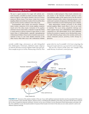
The eye is a good example of an organ with multiple auto- intoxication, is called cyclospasm. Ciliary muscle contraction also
nomic functions, controlled by several autonomic receptors. As puts tension on the trabecular meshwork, opening its pores
shown in Figure 6–9, the anterior chamber is the site of several and facilitating outflow of the aqueous humor into the canal of
autonomic effector tissues. These tissues include three muscles Schlemm. Increased outflow reduces intraocular pressure, a very
(pupillary dilator and constrictor muscles in the iris and the ciliary useful result in patients with glaucoma. All of these effects are pre-
muscle) and the secretory epithelium of the ciliary body. vented or reversed by muscarinic blocking drugs such as atropine.
Parasympathetic nerve activity and muscarinic cholinomi- Alpha adrenoceptors mediate contraction of the radially
metics mediate contraction of the circular pupillary constrictor oriented pupillary dilator muscle fibers in the iris and result
muscle and of the ciliary muscle. Contraction of the pupillary in mydriasis. This occurs during sympathetic discharge and
constrictor muscle causes miosis, a reduction in pupil size. Miosis when α-agonist drugs such as phenylephrine are placed in the
is usually present in patients exposed to large systemic or small conjunctival sac. Beta adrenoceptors on the ciliary epithelium
topical doses of cholinomimetics, especially organophosphate facilitate the secretion of aqueous humor. Blocking these recep-
cholinesterase inhibitors. Ciliary muscle contraction causes accom- tors (with β-blocking drugs) reduces the secretory activity and
modation of focus for near vision. Marked contraction of the reduces intraocular pressure, providing another therapy for
ciliary muscle, which often occurs with cholinesterase inhibitor glaucoma.
currently available drugs: adrenoceptors are easily distinguished greater selectivity may be attainable in the future using drugs that
from cholinoceptors. Furthermore, individual receptor subgroups target post-receptor processes, eg, receptors for second messengers.
can often be selectively activated or blocked within each major type. The next four chapters provide many more examples of this
Some examples are given in the Box: Pharmacology of the Eye. Even useful diversity of autonomic control processes.
Cornea
Canal of Schlemm
Anterior
chamber
Tr abecular meshwork
Dilator (α)
Sphincter (M)
Sclera
Iris
Lens
Ciliary epithelium (β)
Ciliary muscle (M)
FIGURE 6–9 Structures of the anterior chamber of the eye. Tissues with significant autonomic functions and the associated ANS receptors
(α, β, M) are shown in this schematic diagram. Aqueous humor is secreted by the epithelium of the ciliary body, flows into the space in front of
the iris, flows through the trabecular meshwork, and exits via the canal of Schlemm (arrow). Blockade of the β adrenoceptors associated with
the ciliary epithelium causes decreased secretion of aqueous. Blood vessels (not shown) in the sclera are also under autonomic control and
influence aqueous drainage.