
Page 72 - parasitology for medical and clinical laboratoryprofessionals
P. 72
52 CHAPTER 3
Entamoeba histolytica is the most pathogenic of the
intestinal amoeba that plagues humans. Trophozoites of
E. histolytica can sometimes remain in the intestinal lumen
(tubelike opening) for years without causing any damage
and in this case the patients who are asymptomatic are
carriers who can potentially transmit the organisms to
others. The majority (90 percent) of patients fall into this Source: Centers for Disease Control and Prevention (CDC)
group. Asymptomatic carriers are defined as those who
are infected by a given organism but report no symptoms
and show no signs of the condition of amoebiasis. Disease
states in these persons can most often be detected by fe-
cal analyses. The procedure may also reveal cysts of non-
pathogenic E. dispar, which for unknown reasons is not FIGURE 3-10 Entamoeba histolytica cysts that when
invasive or as potentially harmful as those of the pathogenic mature, will reveal four nuclei
E. histolytica (Figure 3-10), which possesses four nuclei.
The nuclei are not always visible at various levels within
the organism so it is necessary to focus up and down at
several levels with the microscope in order to see all the
nuclei present. It is also important to differentiate cysts of
Entamoeba coli that are larger than E. histolytica and have
eight nuclei ( Figure 3-11) from other parasitic organisms,
as the Entamoeba coli organism is also not pathogenic but
may lead to difficulty in the identification of E. histolytica. Source: Centers for Disease Control and Prevention (CDC)
Symptoms
In amoebic colitis the incubation period varies greatly.
During some period of the infection the E. histolytica
organisms may begin to invade the tissues of the intesti-
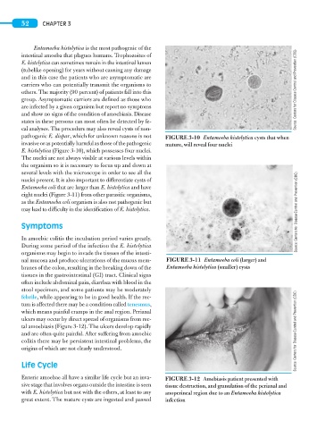
nal mucosa and produce ulcerations of the mucus mem- FIGURE 3-11 Entamoeba coli (larger) and
branes of the colon, resulting in the breaking down of the Entamoeba histolytica (smaller) cysts
tissues in the gastrointestinal (GI) tract. Clinical signs
often include abdominal pain, diarrhea with blood in the
stool specimen, and some patients may be moderately
febrile, while appearing to be in good health. If the rec-
tum is affected there may be a condition called tenesmus,
which means painful cramps in the anal region. Perianal
ulcers may occur by direct spread of organisms from rec-
tal amoebiasis (Figure 3-12). The ulcers develop rapidly
and are often quite painful. After suffering from amoebic Source: Centers for Disease Control and Prevention (CDC)
colitis there may be persistent intestinal problems, the
origins of which are not clearly understood.
Life Cycle
Enteric amoebae all have a similar life cycle but an inva- FIGURE 3-12 Amebiasis patient presented with
sive stage that involves organs outside the intestine is seen tissue destruction, and granulation of the perianal and
with E. histolytica but not with the others, at least to any anoperineal region due to an Entamoeba histolytica
great extent. The mature cysts are ingested and passed infection