
Page 69 - parasitology for medical and clinical laboratoryprofessionals
P. 69
Protozoal Microorganisms as Intestinal Parasites 49
spine or plane of the parasite. Three of these pairs are
attached to the dorsal surface and one pair with a ventral
origin. Some of these flagella, a total of eight, may not be
readily visible microscopically.
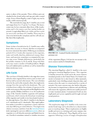
The most infective stage, the G. lamblia cyst, is oval
and ranges from 8 to 17 μm by 7 to 10 μm. The karyo-
somes may be less concentric than those of the tropho-
zoites, and in the mature cyst, four median bodies are
present. Longitudinal fibers are visible and four nuclei
are seen in the cyst form of G. lamblia. There may be a
clear zone between the cytoplasm and the cell wall, un-
like that of the trophozoite. Source: Centers for Disease Control and Prevention (CDC)
Symptoms
Some victims of an infection by G. lamblia may suffer
from either an acute or chronic diarrhea accompanied
by severe intestinal discomfort. Following an incubation
period of 2 to 3 weeks after exposure to the organism, FIGURE 3-8 A stained Giardia intestinalis
water and smelly diarrhea, abdominal cramps, flatu- protozoa cyst
lence, and anorexia, sometimes accompanied by nau-
sea, may occur. Vitamin deficiencies, particularly the of the organisms (Figure 3-8) but the wet mount is the
fat- soluble vitamins A, D, E and K, along with folic most common method of identification.
acid, may create a number of health problems. Weight
loss and malabsorption syndrome as well as steatorrhea Disease Transmission
(fatty stools) may occur.
The intestinal flagellate called G. lamblia is the most
Life Cycle pathogenic intestinal flagellate known. This organism
is familiar around the world and is the most common
The cyst form of Giardia lamblia is the stage that causes intestinal parasite in the United States. It is found com-
infection when ingested from sources such as water and monly in humans but is also found in other mammals,
food as well as several other means. The cyst is broken particularly water-dwelling animals of ponds, lakes, and
down by the acid in the stomach, where the trophozoite streams where the water is contaminated by a variety of
emerges from the cyst wall in the duodenum. The cyst animals. City water systems may also harbor this para-
is broken down within a few minutes of exposure to the site because it is impervious to chlorine and only filtering
acidic environment of the stomach and the flagella become will remove the organisms, which is not readily practiced
active quickly. Within a half hour or so, the trophozoite in water treatment plants. Contaminated foods are also
stage of the Giardia lamblia organism will undergo implicated, such as raw vegetables and through oral-anal
cytokinesis, which restores the binucleate morphology sexual practices.
of the organism and results in two binucleated trophozo-
ites through fission. Note that G. lamblia is also known Laboratory Diagnosis
as G. intestinalis and G. duodenalis. This rapid replica-
tion makes it possible to experience a heavy infection The trophozoite stage of G. lamblia is the most com-
only days following an initial infection. The trophozoite monly found stage and is described as a characteristic
obtains necessary nutrition from the intestinal lumen pear or teardrop shape. These trophozoites are highly
(tube opening) by a process called pinocytosis, where an motile and move in a twisting and erratic manner, sim-
organism or a cell absorbs nutrients and fluids from tis- ilar to that of a falling leaf. These flagella will be more
sue. Giardia is sometimes stained to visualize structures apparent following a review of images of trophozoites