
Page 11 - CJO_F17_GLAUCOMA_SUPPLEMENT
P. 11
MANAGING OPEN ANGLE GLAUCOMA
Identification of risk factors in an individual begins with patient history, while others will be detected by clinical exami-
nation. A number of risk factors for glaucoma have been identified, but it is important to discriminate which of these is
supported by strong evidence. Large prospective longitudinal studies have confirmed the following strong risk factors for
the development of POAG: increased age, elevated IOP, thin central corneal thickness, and increased cup-to-disc ratio. 8,13,58
Race (African-North American, Hispanic heritage) and family history (first-degree relative with glaucoma) are two other
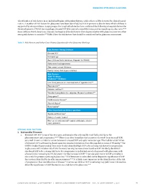
strong risk factors to consider. Table 2 lists the risk factors that should be considered in the glaucoma assessment.
9,59
Table 2: Risk Factors and Other Case History Questions for the Glaucoma Work-up
Risk Factors: Strong Evidence
Elevated IOP
Increased age
Race (African-North American, Hispanic for POAG)
Optic nerve head appearance
Thin central corneal thickness
Family history (first degree relatives)
Risk Factors:
Mild* Evidence
Moderate** Evidence
Low blood pressure (or over-treatment of hypertension)**
High myopia**
Diabetes mellitus**
Vascular dysregulation (i.e. migraine, Raynaud syndrome)**
Sleep apnea**
Cardiovascular disease*
Thyroid (hypo)*
Hypertension*
Other important case history questions
Significant blood loss?
History of ocular trauma?
Prior use of corticosteroid? (topical ophthalmic, inhaled
nasal, systemic)
STRONG RISK FACTORS
1. Intraocular Pressure
a. Elevated IOP: IOP is one of the strongest, and remains the only readily modifiable, risk factor for
glaucoma onset and progression. 12,35,59 There is no clear boundary that separates ‘elevated’ from ‘normal’ IOP,
yet a well-known correlation exists between increased IOP and optic nerve damage. The traditional definition
of elevated IOP is ≥21 mmHg, based upon two standard deviations from the population mean of 15mmHg. The
8
OHTS (Ocular Hypertension Treatment Study) identified high IOP to be a strong risk factor for progression
to glaucoma with relative risk of 10% for every 1mmHg increase above baseline IOP. Similarly, the EMGT
8
also found IOP to be a strong risk factor for progression in individuals newly diagnosed with glaucoma, with
a hazard ratio increasing by 11% for every 1mmHg increase in IOP. Inter-eye asymmetry in IOP ≥2mmHg is
13
also a risk for primary open angle glaucoma. The literature is undecided on the diagnostic significance of IOP
37
fluctuations identified by in-office measurements. 60-62 It is likely more important in the identification of the risk
of progression than identification of disease. Continuous 24-hour IOP monitoring may yield more information
62
about the significance of IOP fluctuation as a risk factor for glaucoma. 63-65
CANADIAN JOURNAL of OPTOMETRY | REVUE CANADIENNE D’OPTOMÉTRIE VOL. 79 SUPPLEMENT 1, 2017 11