
Page 8 - CJO_F17_GLAUCOMA_SUPPLEMENT
P. 8
C CLINICAL RESEARCH
Subsequent examinations must include gonioscopy to further investigate these findings, and to differentially diagnose
POAG, secondary OAG, or ACG. An important part of the anterior segment examination is a careful assessment for con-
current ocular surface disease: its significant impact on management of glaucoma will be reviewed later in this guideline.
Clinical Recommendation for the primary eye care examination:
• A thorough case history, anterior segment exam, and intraocular pressure assessment will identify risk
factors and heighten vigilance for detection of glaucomatous optic neuropathy through clinical assessment
of the optic nerve head complex.
Although elevated intraocular pressure no longer defines glaucoma, it remains one of the most important, and cur-
rently the only readily modifiable risk factor for the disease. Each 1mmHg increase in IOP can increase risk of pro-
33
gression by up to 20%. However, studies have also shown that over half of the patients diagnosed with glaucoma
34
can present with a pressure less than 22mmHg, and a single measurement will miss peak IOP 75% of the time. 35,36
Consistently elevated IOP (≥21mmHg), interocular asymmetry (≥2mmHg), or significant fluctuations particularly
at low IOPs should heighten concern and prompt further investigation. 37,38 Of course, the identification of strong risk
factors and suspicious optic nerve features in the presence of IOP <22mmHg cannot be overlooked.
Systematic clinical assessment of the optic nerve head complex remains the cornerstone of detecting the structural damage
that defines glaucomatous optic neuropathy. If there is a single focus to hang your hat on, this maybe it: retinal ganglion
39
cell loss manifesting as diffuse or focal neuroretinal rim and retinal nerve fiber layer thinning. Optic disc hemorrhages
40
and parapapillary atrophy, often found adjacent to areas of rim loss, are also common signs of the disease. In clinical
41
practice, the cup-to-disc ratio is an easy and efficient value to obtain. However, it is variable between and within observ-
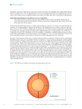
ers, and can be misleading without the context of optic nerve head size. 42-44 The ISNT rule is a quick way to identify the
configuration of a normal optic nerve. As shown in Figure 1, ISNT refers to the rim thickness of a normal optic nerve from
thickest to thinnest: inferior, superior, nasal, temporal. Combining an estimate of the size of the nerve and identification
of whether the ISNT rule is obeyed with the cup-to-disc ratio estimation might be a more sensitive screening evalua-
tion than cup-to-disc ratio alone. 45-47 For example, a cup-to-disc ratio of 0.3 in a small nerve that disobeys the ISNT rule
should be regarded as highly suspicious for glaucoma. Such suspicion should both prompt the clinician to perform a more
thorough evaluation of both the optic nerve and Retinal nerve fiber layer (RNFL) to identify any glaucomatous features
described in the clinical examination section below, and also follow-up with a more comprehensive glaucoma assessment
when clinically indicated.
Figure 1: The ISNT rule as it applies to an average, non-glaucomatous, optic nerve.
8 CANADIAN JOURNAL of OPTOMETRY | REVUE CANADIENNE D’OPTOMÉTRIE VOL. 79 SUPPLEMENT 1, 2017