
Page 13 - CJO_F17_GLAUCOMA_SUPPLEMENT
P. 13
MANAGING OPEN ANGLE GLAUCOMA
4. Family History
a. The inheritance pattern of POAG remains uncertain, but it is accepted that the disease is a complex multifactorial
polygenic disease that commonly manifests in multiple generations of a family. The Rotterdam Eye Study found
71
that the lifetime risk of developing POAG at 80 years of age was 10 times higher in individuals with a family
history of glaucoma than in those without. 72
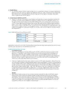
5. Central Corneal Thickness (CCT)
a. Thin CCT (< 555 um): A thin central corneal thickness was found to be a strong and independent risk factor for
conversion from OHT (ocular hypertension) to POAG in the OHTS. In fact, in a multivariate analysis of all
8,73
the significant risk factors for progression from OHT to POAG, CCT was the strongest with a relative risk ratio
of 70% for every 40μm decrease in thickness from baseline. The results from this model are adapted and shown
below in Table 3. When considering relative risk for glaucoma, rather than adjusting IOP to correct for CCT, it is
more valuable to simply classify CCT as thin, average or thick. 73,74 It is still unclear as to whether thin CCT is only
a predictor for progression to glaucoma from OHT, or if it is also a risk factor for progression once glaucoma has
been diagnosed. 8,58,75
Table 3: OHTS and Central Corneal Thickness (CCT)
IOP>25.75 36% 13% 6%
IOP> 23.75 12% 10% 7%
17% 9% 2%
IOP < 23.75
CCT <555 >555 to< 588 >588
Adapted from: Gordon, M. O., et al. (2002) The Ocular Hypertension Treatment Study: baseline factors that predict the onset
of primary open-angle glaucoma, Arch Ophthalmol, 120, 714-20. 8
OTHER IMPORTANT RISK FACTORS
Low Blood Pressure:
• There is an established link between POAG, specifically NTG, and low blood pressure and poor ocular
blood flow. 29,61 The EMGT found that low blood pressure was an important risk factor for progression
among subjects with glaucoma regardless of baseline IOP. A patient might have low blood pressure
76
physiologically, or as a result of over treatment for systemic hypertension. If a patient being evaluated for
glaucoma is being treated for high blood pressure it is important to identify the type and dosage of the
medication, as well as the time of day it is administered. 77
• It has been hypothesized that low ocular perfusion pressure (OPP) leads to alterations in blood flow at the
optic nerve and contributes to progressive glaucomatous optic nerve damage. 29,76 Diastolic ocular perfusion
pressure (DOPP) can be quickly estimated in the clinical setting to identify individuals who likely have low
vascular perfusion to the optic nerve. This simple estimation involves taking the difference of the diastolic
blood pressure (DBP) and IOP (DOPP = DBP - IOP). The Baltimore Eye Survey found that low DOPP was
strongly associated with the prevalence of glaucoma. 12,29 It has been suggested that DOPP values of less
than 56 can be a useful threshold to identify patients at increased risk of progressive glaucomatous optic
neuropathy. 78
Myopia:
• High myopia: Various studies and meta-analyses have demonstrated that subjects with higher myopic
refractive error have a significantly greater prevalence of glaucoma than groups with low myopia or
emmetropia. 79,80 This association exists as a risk factor for both development and progression of POAG.
The underlying hypothesis is that individuals with greater axial length accompanying high myopia have
weaker scleral support for retinal ganglion cells at the lamina cribrosa and this weakness increases the
susceptibility of the optic nerve to glaucomatous damage.
31
CANADIAN JOURNAL of OPTOMETRY | REVUE CANADIENNE D’OPTOMÉTRIE VOL. 79 SUPPLEMENT 1, 2017 13