
Page 33 - CJO_F17_FLIPBOOK
P. 33
RESEARCH
modalities. However, due to the rarity of the condition, little information exists to determine long-term prognosis.
The condition is a chronic one, and does require continued monitoring and possibly long-term treatment.
Vision loss from Type II IJT develops due to atrophy of the outer retina and photoreceptors. Due to the location
of retinal atrophy being temporal to the foveal center, visual acuity often remains good in these patients until
the development of SRNV. Data from the MacTel Study group showed that 42% of all patients had visual acuity
of 20/25 or better. However, this photoreceptor damage can create paracentral scotomas that lead to patient
34
symptoms even with good retained visual acuity. Various reports demonstrate that even with acceptable visual
35
acuity, patients become symptomatic for patient perceived metamorphopsia, decreased reading speed, and dif-
ficulty in reading – the latter of which being the most frequently reported initial symptom. 36,37 These symptoms
arise most commonly between the ages of 50 and 69. Exact values for long-term prognosis for Type II IJT varies
between reports but patients do have increased risk for both decreased visual acuity as well as decreased quality
of life from vision loss. 38,39
CONCLUSION
Diagnosis of IJT can be complicated by clinical appearances mimicking other conditions. For example Type I IJT
can easily be confused with diabetic retinal changes. While treatment options for the complication of macular ede-
ma from Type I IJT are essentially the same as those for diabetic macular edema, it is still important to make the
correct diagnosis. While systemic conditions should still be considered and ruled out, a patient with Type I IJT
could have unwarranted psychological distress if told they have retinal complications from diabetes when they
don’t have the disease.
Also, even though there is currently no known beneficial treatment in the case of Type II IJT without SRNV,
it is still important to make an accurate assessment in order to educate patients properly about their condition
and prognosis. Patients should be made aware that the condition is a progressive retinal disease which can lead
to central vision loss, metamorphopsia, and paracentral scotomas. They also need to be thoroughly educated
on how to properly monitor their vision monocularly, and to be examined periodically for SRNV. With proper
understanding of IJT, optometrists can monitor these conditions appropriately, referring for additional treat-
ment when necessary. l
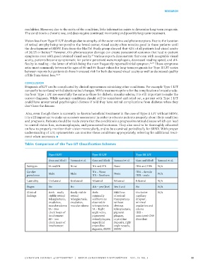
Table: Comparison of the Two IJT Classification Schemes
Type I IJT Type II IJT Type III IJT
Gass and Blodi Yanuzzi et al. Gass and Blodi Yanuzzi et al. Gass and Blodi Yanuzzi et al.
Subtypes IA and IB None IIA and IIB None IIIA and IIIB N/A
Gender IIA – None IIIA – female
prevalence Male Male IIB – Brothers None IIIB – male N/A
Laterality Unilateral Unilateral Bilateral Bilateral Bilateral N/A
Stages No No IIA – yes (five) Yes (two) No N/A
Clinical Both – easily Easily visible Both – Mild loss Occlusive N/A
findings visible retinal retinal minimally of retinal capillary
telangiectasia, telangiectasia, visible to no transperancy dropout,
exudation, exudation, observable without minimal
macular edema macular edema telangectiasia obvious exudation and
IA > two IA – Right telangiectasia, edema
clock hours of angle veins, pigment IIIB –
involvement pigmented plaques, associated CNS
IB < two retinal plaques, crystalline disorders
clock hours of superficial deposits, right
involvement crystalline angle vessels,
deposits, SRNV SRNV
CANADIAN JOURNAL of OPTOMETRY | REVUE CANADIENNE D’OPTOMÉTRIE VOL. 79 NO. 3 33