
Page 31 - CJO_F17_FLIPBOOK
P. 31
RESEARCH
Type II IJT may be an acquired condition in which the malformations of the retinal vasculature present later in
life. A histological study performed by Green et al. on a patient with Type II IJT showed narrowing of the vessel
3,4
lumen versus the telangiectatic or dilated appearance noted clinically and on FA. Increased width of the basement
membrane along with degeneration of pericytes and endothelial cells led to thickening of the retinal capillaries.
The authors conceived that breaks in the endothelium allowed fluorescein to leak into the thickened capillary walls
which accounted for the late staining seen on FA. This report also found that capillary proliferation extended into
the outer retinal layers down to the photoreceptors. A different histopathological report from a more advanced
10
case of Type II IJT with SRNV did show dilation of retinal capillaries that reached into the outer retinal layers and
subretinal space. Pigment migration also occurred along the course of the proliferating vessels. 11
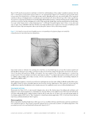
Figure 7: FAF details an area of central hypofluorescence corresponding to the pigment plaque surrounded by
a hyperfluorescent ring indicating tissue degradation.
Speculation exists on whether the capillary abnormalities are the initial step in a process that leads to nutritional
and metabolic damage to the retina, or if there is some sort of retinal change that precedes and leads to the forma-
tion of the abnormal capillaries. Müller cells garner the most suspicion due to their importance in maintaining
proper health of the vasculature endothelium and sensory retina. 12,13 A primary dysfunction in these cells may lead
to capillary abnormalities and retinal atrophy, with the superficial crystalline deposits representing remnants of
degenerated Müller cells. 14
SRNV formation appears to stem from proliferation and anastomoses that exist within the blood vessels of the retina.
This differs from the retino-choroidal anastomoses found in macular degeneration. The reason for retinal capillary
proliferation is unclear, but photoreceptor atrophy may give the capillaries easier access to the subretinal space.
15
TREATMENT OPTIONS
Treatments for Type I IJT, or aneurysmal telangiectasia, aim at the destruction of the abnormal capillaries and
aneurysms with photocoagulation or photodynamic therapy (PDT); or the stabilization of the blood retinal barrier
with injectable medications. Limited options exist for those with Type II IJT due to the non-edematous nature of
the disease, unless SRNV forms. Then treatments include laser photocoagulation, PDT, intravitreal triamcinolone
injections, and intravitreal anti VEG-F agents.
Photocoagulation
Photocoagulation benefitted only those with Type I IJT, as noted by both Gass and Owakawa; and Gass and Blodi in
their respective reports. They found a potential improvement in vision and exudation with photocoagulation. The
procedure proved to be non-beneficial in those with Type II IJT. 3,16
CANADIAN JOURNAL of OPTOMETRY | REVUE CANADIENNE D’OPTOMÉTRIE VOL. 79 NO. 3 31