
Page 26 - CJO_F17_FLIPBOOK
P. 26
C CLINICAL RESEARCH
INTRODUCTION
IJT is a condition comprised of three classical divisions, each with different clinical findings, etiologies, and patient
demographics. They do share a common feature of capillary abnormalities in the juxtafoveal region that appear on
fluorescein angiography as dilated or telangiectatic capillary changes. IJT can easily be confused for other condi-
1
tions, and these abnormal capillary findings must be separated from those resultant from other conditions such as
diabetes, carotid occlusive disease, retinal vein occlusion, radiation therapy, and others. The classification also does
2
not include Coat’s disease, which has more widespread retinal telangiectatic changes. However, Coat’s disease and
IJT Type I could be subsets of the same condition that exists as a spectrum of disease. 3
GASS AND BLODI CLASSIFICATION
Gass and Owakawa first coined the term IJT in 1982 and initially categorized the condition in four different
classes. Gass and Blodi later revisited the topic in 1993 in a follow up report of the initial work done by Gass and
1
Owakawa at which time they modified the classification into three main categories, each with separate unrelated
etiologies and two subsets: Type IA and B, Type IIA and B, and Type IIIA and B. Additionally, Type II was further
divided into five stages. Clinical observations and fluorescein angiography (FA) findings formed the basis of the
3
grouping in this system.
Type I
Type I IJT presented with easily visible retinal telangiectasia often accompanied with exudation and macular ede-
ma, with vision loss resulting from the latter. Gass and Blodi broke down Type I IJT based on the extent of telangi-
ectasia as defined by the number of involved clock hours around the fovea. Type IA had more than two clock hours
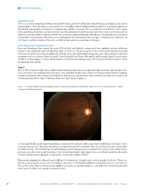
of telangiectasia while Type IB had less than two clock hours (Figure 1).
Figure 1: Easily visible retinal telangiectasia in a patient with Gass and Blodi Type IA IJT, or Yanuzzi et al.’s Type I
aneurysmal telangiectasia.
In Gass and Blodi’s study, Type IA patients consisted of 31 subjects with a high male predilection (28 male) and a
mean patient age of 37. Entering visual acuity ranged from 20/20 to 20/200. They found Type IA to be unilateral in
30 of the 31 cases. Clinical findings varied between patients, but generally presented with easily visible retinal telan-
giectasia, macular edema, and exudation. Type IA patients lacked right angle veins, superficial crystalline deposits,
intraretinal pigmentary plaques, or subretinal neovascularization (SRNV).
Fluorescein angiography showed quick filling of the irregular telangiectatic vessels around the fovea. These ap-
peared as an irregular round zone of capillary aneurysms and dilated capillaries located temporal to the fovea in
94% of the cases. They noted minimal capillary occlusion or capillary dropout. Late stage FA revealed intraretinal
staining as the abnormal vessels continued to leak.
26 CANADIAN JOURNAL of OPTOMETRY | REVUE CANADIENNE D’OPTOMÉTRIE VOL. 79 NO. 3