
Page 27 - CJO_F17_FLIPBOOK
P. 27
RESEARCH
Only eight patients in the study displayed the less prevalent Type IB IJT. Again, a high gender predilection existed with
seven of the patients being male. The average age was 42. All but two eyes (20/25 and 20/30) exhibited 20/20 visual acuity.
Findings were again unilateral in most cases (seven out of eight) but were confined to two clock hours or less of still
easily visible telangiectasia. A few flecks of exudates occurred in most cases, but less than that seen with Type IA.
Fluorescein angiography showed early filling of one to four capillary aneurysms and of the irregular capillaries fol-
lowed by late staining around the rare aneurysms.
Type II
Unlike Type I, Type II IJT rarely displays macular edema and exudation. In these patients, outer retina atrophy is
the main cause of vision loss. Gass and Blodi classified Type II into subsets A and B based on the age of presentation.
Type IIA consisted of 92 patients with equal gender incidence and an average age of 55 years. Entering visual
acuity ranged from 20/15 to hand motion. Presentation was bilateral, though often asymmetric, in 90 out of
92 patients. Unlike Type I, these patients exhibited either minimal, poorly visible, or no clinically discernible
telangiectasia. These abnormal capillaries would light up early with FA and then gradually stain in late stages
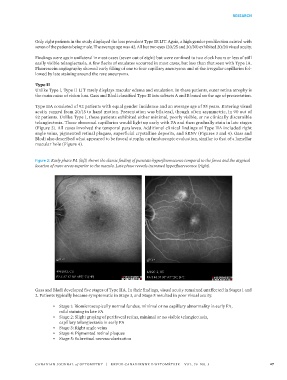
(Figure 2). All cases involved the temporal parafovea. Additional clinical findings of Type IIA included right
angle veins, pigmented retinal plaques, superficial crystalline deposits, and SRNV (Figures 3 and 4). Gass and
Blodi also described what appeared to be foveal atrophy on funduscopic evaluation, similar to that of a lamellar
macular hole (Figure 4).
Figure 2: Early phase FA (left) shows the classic finding of punctate hyperfluorescence temporal to the fovea and the atypical
location of more areas superior to the macula. Late phase reveals increased hyperfluorescence (right).
Gass and Blodi developed five stages of Type IIA. In their findings, visual acuity remained unaffected in Stages 1 and
2. Patients typically became symptomatic in Stage 3, and Stage 5 resulted in poor visual acuity.
• Stage 1: Biomicroscopically normal fundus, minimal or no capillary abnormality in early FA,
mild staining in late FA
• Stage 2: Slight graying of perifoveal retina, minimal or no visible telangiectasia,
capillary telangiectasia in early FA
• Stage 3: Right angle veins
• Stage 4: Pigmented retinal plaques
• Stage 5: Subretinal neovascularization
CANADIAN JOURNAL of OPTOMETRY | REVUE CANADIENNE D’OPTOMÉTRIE VOL. 79 NO. 3 27