
Page 28 - CJO_F17_FLIPBOOK
P. 28
C CLINICAL RESEARCH
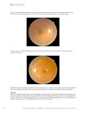
Figure 3: Gass and Blodi Type IIA Stage 4 (note the prominent right angle vein stretching from the superior temporal venous
branch directly to the pigment plaque), or Yanuzzi et al.’s Type II perifoveal telangiectasia, non-proliferative stage.
Figure 4: Type II IJT with crystalline deposits, perifoveal retinal whitening, and a foveal appearance similar to that of a
lamellar macular hole.
Type IIB consisted of only two patients who were brothers aged nine and 12. Both presented with bilateral SRNV
and subtle juxtafoveal telangiectasia, but without right angle veins, crystalline deposits, or pigmented plaques.
Type III
Occlusive capillary dropout along with retinal telangiectasia characterized Type III. All had minimal exudation and
edema. Type IIIA consisted of three women aged 41 to 59, all with bilateral findings. Their visual acuity ranged from
20/20 to 20/50. One reported polycythemia vera, another gout, and a third hyperglycemia. Type IIIB consisted of
five men aged 35 to 41 all with CNS disorders. Two of them were brothers.
28 CANADIAN JOURNAL of OPTOMETRY | REVUE CANADIENNE D’OPTOMÉTRIE VOL. 79 NO. 3