
Page 10 - SPRING 2016
P. 10
Digital Dental Smile
By Benjamin Schwartz, DDS, FAGD, FICOI
Congratulations! You’ve earned your DDS diploma from an DIGITAL SHADE MACHINES
accredited dental school! Whether it be last month or 25 years If you’ve ever had to return a case to the lab for a shade modifi-
ago, you are proud of your accomplishment, but are you prepared cation, you understand how frustrating and time consuming that
to earn your Digital Dental Smile diploma as well? is. By utilizing a digital shade machine, this headache can be a
thing of the past. It quickly and accurately determines the correct
Each day in the field of dental technology, more and more shade of the tooth so that color matches are no longer a guessing
advances occur. If you’re not keeping pace with these improve- game.
ments, you’ll certainly be left behind!
CARIES DETECTORS
The purpose of this article is to highlight some of the innovations Often times, carious lesions can be difficult to detect, even on a
that are readily available today and their integration into a gener- radiograph. A caries detection system helps to identify decay,
al dental practice. especially in teeth that may otherwise look healthy. In addition,
you won’t need to ‘watch’ teeth, as the systems provide a diag-
DIGITAL RADIOGRAPHS nostic numerical readout that can easily be checked at each recare
By now, most offices are on board with this technology. Its many appointment.
advantages include its ease of use, no more harsh chemicals,
lower radiation dosage, significant time savings, lower cost, and 3-D PRINTERS
the ability to enhance images. A recent addition to the digital dental field, these are fast becom-
ing a mainstay. With the ability to print surgical implant guides,
CONE BEAM COMPUTED TOMOGRAPHY custom trays, night guards, sports guards and retainers, their ver-
For offices that perform dental implants, third molar removal, and satility and accuracy are unparalleled.
endodontic procedures, this item is a must have. The ability to
view teeth and vital structures in three dimensions, while accu- Below are a series of example cases whereby utilizing some of
rately measuring distances or widths, is crucial to a successful the digital dental smile technology, we were able to improve our
treatment outcome. patients’ dental health.
DIGITAL IMPRESSION SYSTEM DIGITAL DENTAL IMPLANT
®
With the advent of CEREC technology over 30 years ago, the A 26-year-old female presented with retained primary tooth #A.
concept of needing to take a physical impression has been upend- The tooth had recently started bothering her and was very sensi-
ed. By taking a digital impression, there is no need for gloopy, tive while eating on it. An intra-oral examination revealed the pri-
gag-inducing material to be shoved into patients’ mouths any mary tooth with recession on the mesial aspect and a digital radi-
longer! You can design virtual wax-ups, crowns, bridges, veneers, ograph (Schick 33, Sirona Dental) was taken, which showed a
night guards, implant restorations, and even dentures all from a large area of decay. After a thorough discussion with the patient,
digital impression. which included intra-oral images (Figures 1, 2) (Iris Camera,
Digital Doc), the treatment option of extraction and immediate
IN-OFFICE MILLING UNITS
implant placement with a delayed restoration was chosen by the
The ability to mill permanent restorations in-office is a definite patient.
game changer. No longer do patients need to wear temporaries for
weeks on end while the dental laboratory fabricates their final A full dental workup was performed on the patient, including a
prosthesis. No additional appointments are needed for insertion, digital impression of the arch utilizing the CEREC software
®
as everything can be accomplished in just one visit. (Sirona Dental) and a cone beam radiograph (Orthophos XG-3D,
Sirona Dental) for implant planning. With the use of Blue Sky
INTRA-ORAL CAMERAS Plan CBCT software (Figure 3) (Blue Sky Bio), a digital implant
By taking an actual photograph of the affected area, the dentist guide was designed and sent to a 3-D printer (Stratasys, A full dental workup was performed on the patient, including a
and patient are able to have a discussion about the issue. Patients Digi3DWorks) for fabrication.
readily point out problem areas on their teeth and help to co-diag- d
nose their own conditions.
N Given that the entire
Figure 3. Digital
p
A full dental workup was performed on the patient, including a
planning of
d implant
placement.
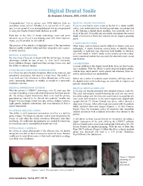
Figures 1 and 2. Pre-operative view of tooth A.
www.nysagd.org | Spring 2017 | GP 10
A full dental workup was performed on the patient, including a
d N Given that the entire
p
N Given that the entire
p